OpenStax College, OpenStax College
Chapter 18. Animal Reproduction and Development
Introduction*
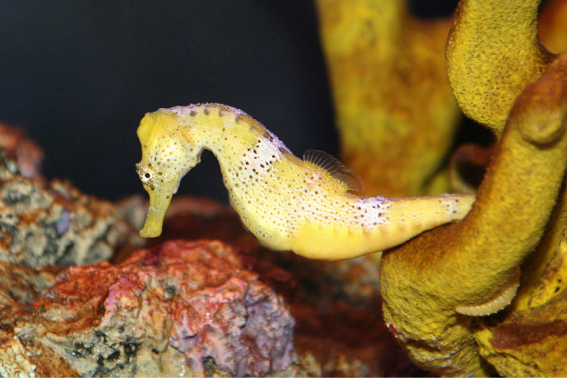
In the animal kingdom, each species has its unique adaptations for reproduction. Asexual reproduction produces genetically identical offspring (clones), whereas in sexual reproduction, the genetic material of two individuals combines to produce offspring that are genetically different from their parents. During sexual reproduction the male gamete (sperm) may be placed inside the female’s body for internal fertilization, the sperm may be left in the environment for the female to pick up and place in her body, or both sperm and eggs may be released into the environment for external fertilization. Seahorses provide an example of the latter, but with a twist (Figure 18.1). Following a mating dance, the female releases eggs into the male seahorse’s abdominal brood pouch and the male releases sperm into the water, which then find their way into the brood pouch to fertilize the eggs. The fertilized eggs develop in the pouch for several weeks.
18.1. How Animals Reproduce*
By the end of this section, you will be able to:
- Describe advantages and disadvantages of asexual and sexual reproduction
- Discuss asexual reproduction methods
- Discuss sexual reproduction methods
- Discuss internal and external methods of fertilization
Some animals produce offspring through asexual reproduction while other animals produce offspring through sexual reproduction. Both methods have advantages and disadvantages. Asexual reproduction produces offspring that are genetically identical to the parent because the offspring are all clones of the original parent. A single individual can produce offspring asexually and large numbers of offspring can be produced quickly; these are two advantages that asexually reproducing organisms have over sexually reproducing organisms. In a stable or predictable environment, asexual reproduction is an effective means of reproduction because all the offspring will be adapted to that environment. In an unstable or unpredictable environment, species that reproduce asexually may be at a disadvantage because all the offspring are genetically identical and may not be adapted to different conditions.
During sexual reproduction, the genetic material of two individuals is combined to produce genetically diverse offspring that differ from their parents. The genetic diversity of sexually produced offspring is thought to give sexually reproducing individuals greater fitness because more of their offspring may survive and reproduce in an unpredictable or changing environment. Species that reproduce sexually (and have separate sexes) must maintain two different types of individuals, males and females. Only half the population (females) can produce the offspring, so fewer offspring will be produced when compared to asexual reproduction. This is a disadvantage of sexual reproduction compared to asexual reproduction.
Asexual Reproduction
Asexual reproduction occurs in prokaryotic microorganisms (bacteria and archaea) and in many eukaryotic, single-celled and multi-celled organisms. There are several ways that animals reproduce asexually, the details of which vary among individual species.
Fission
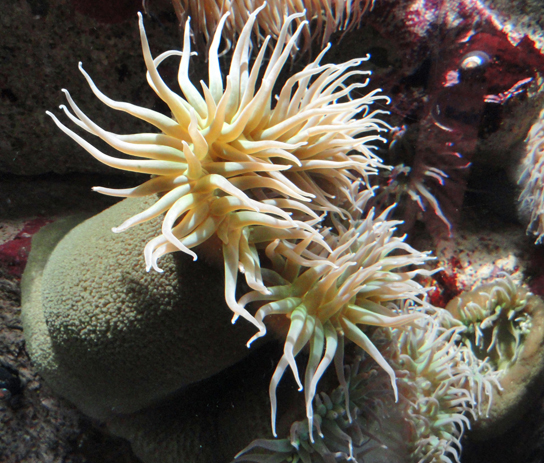
Fission, also called binary fission, occurs in some invertebrate, multi-celled organisms. It is in some ways analogous to the process of binary fission of single-celled prokaryotic organisms. The term fission is applied to instances in which an organism appears to split itself into two parts and, if necessary, regenerate the missing parts of each new organism. For example, species of turbellarian flatworms commonly called the planarians, such as Dugesia dorotocephala, are able to separate their bodies into head and tail regions and then regenerate the missing half in each of the two new organisms. Sea anemones (Cnidaria), such as species of the genus Anthopleura (Figure 18.2), will divide along the oral-aboral axis, and sea cucumbers (Echinodermata) of the genus Holothuria, will divide into two halves across the oral-aboral axis and regenerate the other half in each of the resulting individuals.
Budding
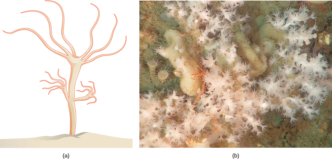
Budding is a form of asexual reproduction that results from the outgrowth of a part of the body leading to a separation of the “bud” from the original organism and the formation of two individuals, one smaller than the other. Budding occurs commonly in some invertebrate animals such as hydras and corals. In hydras, a bud forms that develops into an adult and breaks away from the main body (Figure 18.3).

View this video to see a hydra budding.
Fragmentation
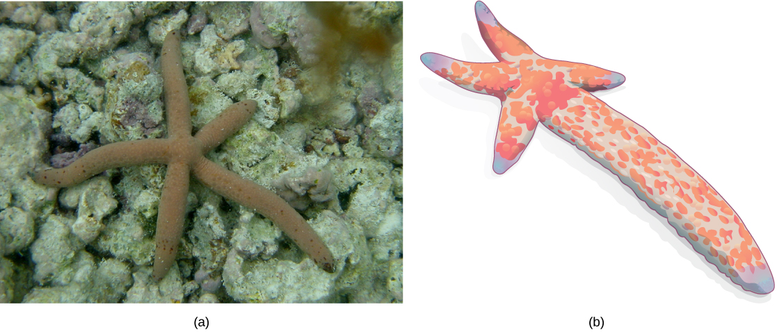
Fragmentation is the breaking of an individual into parts followed by regeneration. If the animal is capable of fragmentation, and the parts are big enough, a separate individual will regrow from each part. Fragmentation may occur through accidental damage, damage from predators, or as a natural form of reproduction. Reproduction through fragmentation is observed in sponges, some cnidarians, turbellarians, echinoderms, and annelids. In some sea stars, a new individual can be regenerated from a broken arm and a piece of the central disc. This sea star (Figure 18.4) is in the process of growing a complete sea star from an arm that has been cut off. Fisheries workers have been known to try to kill the sea stars eating their clam or oyster beds by cutting them in half and throwing them back into the ocean. Unfortunately for the workers, the two parts can each regenerate a new half, resulting in twice as many sea stars to prey upon the oysters and clams.
Parthenogenesis
Parthenogenesis is a form of asexual reproduction in which an egg develops into an individual without being fertilized. The resulting offspring can be either haploid or diploid, depending on the process in the species. Parthenogenesis occurs in invertebrates such as water fleas, rotifers, aphids, stick insects, and ants, wasps, and bees. Ants, bees, and wasps use parthenogenesis to produce haploid males (drones). The diploid females (workers and queens) are the result of a fertilized egg.
Some vertebrate animals—such as certain reptiles, amphibians, and fish—also reproduce through parthenogenesis. Parthenogenesis has been observed in species in which the sexes were separated in terrestrial or marine zoos. Two female Komodo dragons, a hammerhead shark, and a blacktop shark have produced parthenogenic young when the females have been isolated from males. It is possible that the asexual reproduction observed occurred in response to unusual circumstances and would normally not occur.
Sexual Reproduction
Sexual reproduction is the combination of reproductive cells from two individuals to form genetically unique offspring. The nature of the individuals that produce the two kinds of gametes can vary, having for example separate sexes or both sexes in each individual. Sex determination, the mechanism that determines which sex an individual develops into, also can vary.
Hermaphroditism
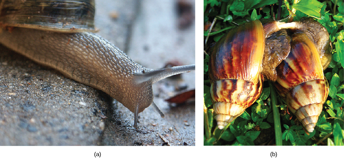
Hermaphroditism occurs in animals in which one individual has both male and female reproductive systems. Invertebrates such as earthworms, slugs, tapeworms, and snails (Figure 18.5) are often hermaphroditic. Hermaphrodites may self-fertilize, but typically they will mate with another of their species, fertilizing each other and both producing offspring. Self-fertilization is more common in animals that have limited mobility or are not motile, such as barnacles and clams. Many species have specific mechanisms in place to prevent self-fertilization, because it is an extreme form of inbreeding and usually produces less fit offspring.
Sex Determination
Mammalian sex is determined genetically by the combination of X and Y chromosomes. Individuals homozygous for X (XX) are female and heterozygous individuals (XY) are male. In mammals, the presence of a Y chromosome causes the development of male characteristics and its absence results in female characteristics. The XY system is also found in some insects and plants.
Bird sex determination is dependent on the combination of Z and W chromosomes. Homozygous for Z (ZZ) results in a male and heterozygous (ZW) results in a female. Notice that this system is the opposite of the mammalian system because in birds the female is the sex with the different sex chromosomes. The W appears to be essential in determining the sex of the individual, similar to the Y chromosome in mammals. Some fish, crustaceans, insects (such as butterflies and moths), and reptiles use the ZW system.
More complicated chromosomal sex determining systems also exist. For example, some swordtail fish have three sex chromosomes in a population.
The sex of some other species is not determined by chromosomes, but by some aspect of the environment. Sex determination in alligators, some turtles, and tuataras, for example, is dependent on the temperature during the middle third of egg development. This is referred to as environmental sex determination, or more specifically, as temperature-dependent sex determination. In many turtles, cooler temperatures during egg incubation produce males and warm temperatures produce females, while in many other species of turtles, the reverse is true. In some crocodiles and some turtles, moderate temperatures produce males and both warm and cool temperatures produce females.
Individuals of some species change their sex during their lives, switching from one to the other. If the individual is female first, it is termed protogyny or “first female,” if it is male first, it is termed protandry or “first male.” Oysters are born male, grow in size, and become female and lay eggs. The wrasses, a family of reef fishes, are all sequential hermaphrodites. Some of these species live in closely coordinated schools with a dominant male and a large number of smaller females. If the male dies, a female increases in size, changes sex, and becomes the new dominant male.
Fertilization
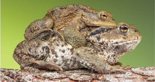
The fusion of a sperm and an egg is a process called fertilization. This can occur either inside ( internal fertilization) or outside ( external fertilization) the body of the female. Humans provide an example of the former, whereas frog reproduction is an example of the latter.
External Fertilization
External fertilization usually occurs in aquatic environments where both eggs and sperm are released into the water. After the sperm reaches the egg, fertilization takes place. Most external fertilization happens during the process of spawning where one or several females release their eggs and the male(s) release sperm in the same area, at the same time. The spawning may be triggered by environmental signals, such as water temperature or the length of daylight. Nearly all fish spawn, as do crustaceans (such as crabs and shrimp), mollusks (such as oysters), squid, and echinoderms (such as sea urchins and sea cucumbers). Frogs, corals, mayflies, and mosquitoes also spawn (Figure 18.6).
Internal Fertilization
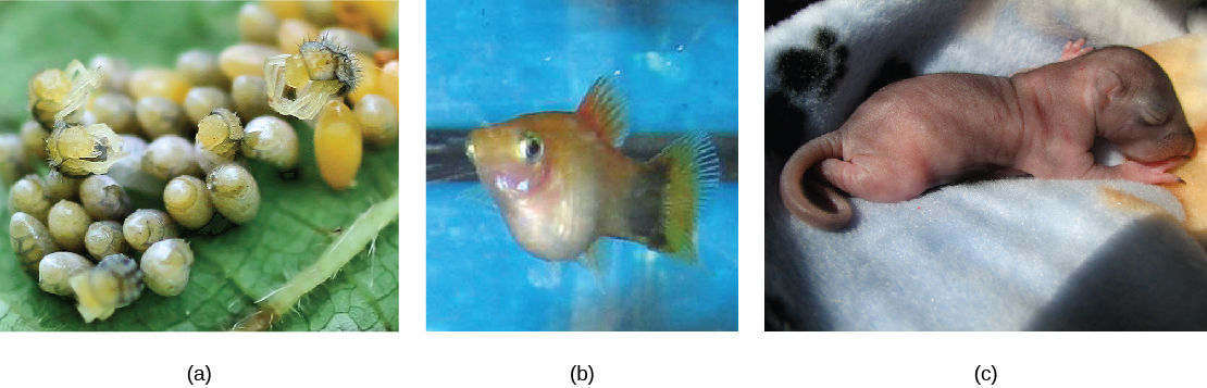
Internal fertilization occurs most often in terrestrial animals, although some aquatic animals also use this method. Internal fertilization may occur by the male directly depositing sperm in the female during mating. It may also occur by the male depositing sperm in the environment, usually in a protective structure, which a female picks up to deposit the sperm in her reproductive tract. There are three ways that offspring are produced following internal fertilization. In oviparity, fertilized eggs are laid outside the female’s body and develop there, receiving nourishment from the yolk that is a part of the egg (Figure 18.7a). This occurs in some bony fish, some reptiles, a few cartilaginous fish, some amphibians, a few mammals, and all birds. Most non-avian reptiles and insects produce leathery eggs, while birds and some turtles produce eggs with high concentrations of calcium carbonate in the shell, making them hard. Chicken eggs are an example of a hard shell. The eggs of the egg-laying mammals such as the platypus and echidna are leathery.
In ovoviparity, fertilized eggs are retained in the female, and the embryo obtains its nourishment from the egg’s yolk. The eggs are retained in the female’s body until they hatch inside of her, or she lays the eggs right before they hatch. This process helps protect the eggs until hatching. This occurs in some bony fish (like the platyfish Xiphophorus maculatus, Figure 18.7b), some sharks, lizards, some snakes (garter snake Thamnophis sirtalis), some vipers, and some invertebrate animals (Madagascar hissing cockroach Gromphadorhina portentosa).
In viviparity the young are born alive. They obtain their nourishment from the female and are born in varying states of maturity. This occurs in most mammals (Figure 18.7c), some cartilaginous fish, and a few reptiles.
18.2. Development and Organogenesis*
By the end of this section, you will be able to:
- Explain how the embryo forms from the zygote
- Discuss the role of cleavage and gastrulation in animal development
- Describe organogenesis
The process by which an organism develops from a single-celled zygote to a multi-cellular organism is complex and well regulated. The regulation occurs through signaling between cells and tissues and responses in the form of differential gene expression.
Early Embryonic Development
Fertilization is the process in which gametes (an egg and sperm) fuse to form a zygote (Figure 18.8). To ensure that the offspring has only one complete diploid set of chromosomes, only one sperm must fuse with one egg. In mammals, a layer called the zona pellucida protects the egg. At the tip of the head of a sperm cell is a structure like a lysosome called the acrosome, which contains enzymes. When a sperm binds to the zona pellucida, a series of events, called the acrosomal reactions, take place. These reactions, involving enzymes from the acrosome, allow the sperm plasma membrane to fuse with the egg plasma membrane and permit the sperm nucleus to transfer into the ovum. The nuclear membranes of the egg and sperm break down and the two haploid nuclei fuse to form a diploid nucleus or genome.
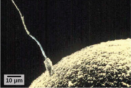
To ensure that no more than one sperm fertilizes the egg, once the acrosomal reactions take place at one location of the egg membrane, the egg releases proteins in other locations to prevent other sperm from fusing with the egg.
The development of multi-cellular organisms begins from this single-celled zygote, which undergoes rapid cell division, called cleavage (Figure 18.9a), to form a hollow ball of cells called a blastula (Figure 18.9b).
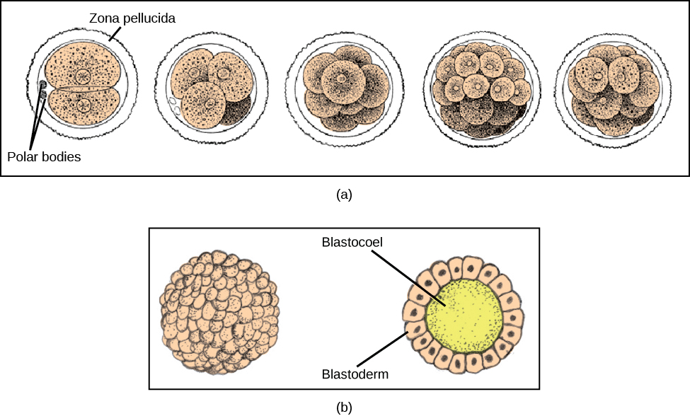
In mammals, the blastula forms the blastocyst in the next stage of development. Here the cells in the blastula arrange themselves in two layers: the inner cell mass, and an outer layer called the trophoblast. The inner cell mass will go on to form the embryo. The trophoblast secretes enzymes that allow implantation of the blastocyst into the endometrium of the uterus. The trophoblast will contribute to the placenta and nourish the embryo.

Visit the Virtual Human Embryo project at the Endowment for Human Development site to click through an interactive of the stages of embryo development, including micrographs and rotating 3-D images.
The cells in the blastula then rearrange themselves spatially to form three layers of cells. This process is called gastrulation. During gastrulation, the blastula folds in on itself and cells migrate to form the three layers of cells (Figure 18.10) in a structure, the gastrula, with a hollow space that will become the digestive tract. Each of the layers of cells is called a germ layer and will differentiate into different organ systems.
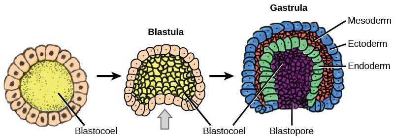
The three germ layers are the endoderm, the ectoderm, and the mesoderm. Cells in each germ layer differentiate into tissues and embryonic organs. The ectoderm gives rise to the nervous system and the epidermis, among other tissues. The mesoderm gives rise to the muscle cells and connective tissue in the body. The endoderm gives rise to the gut and many internal organs.
Organogenesis
Gastrulation leads to the formation of the three germ layers that give rise during further development to the different organs in the animal body. This process is called organogenesis.
Organs develop from the germ layers through the process of differentiation. During differentiation, the embryonic stem cells express specific sets of genes that will determine their ultimate cell type. For example, some cells in the ectoderm will express the genes specific to skin cells. As a result, these cells will take on the shape and characteristics of epidermal cells. The process of differentiation is regulated by location-specific chemical signals from the cell’s embryonic environment that sets in play a cascade of events that regulates gene expression.
18.3. Human Reproduction*
By the end of this section, you will be able to:
- Describe human male and female reproductive anatomies
- Describe spermatogenesis and oogenesis and discuss their differences and similarities
- Describe the role of hormones in human reproduction
- Describe the roles of male and female reproductive hormones
As in all animals, the adaptations for reproduction in humans are complex. They involve specialized and different anatomies in the two sexes, a hormone regulation system, and specialized behaviors regulated by the brain and endocrine system.
Human Reproductive Anatomy
The reproductive tissues of male and female humans develop similarly in utero until about the seventh week of gestation when a low level of the hormone testosterone is released from the gonads of the developing male. Testosterone causes the primitive gonads to differentiate into male sexual organs. When testosterone is absent, the primitive gonads develop into ovaries. Tissues that produce a penis in males produce a clitoris in females. The tissue that will become the scrotum in a male becomes the labia in a female. Thus the male and female anatomies arise from a divergence in the development of what were once common embryonic structures.
Male Reproductive Anatomy
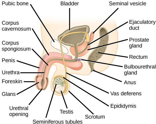
Sperm are immobile at body temperature; therefore, the testes are external to the body so that a correct temperature is maintained for motility. In land mammals, including humans, the pair of testes must be suspended outside the body so the environment of the sperm is about 2 °C lower than body temperature to produce viable sperm. If the testes do not descend through the abdominal cavity during fetal development, the individual has reduced fertility.
The scrotum houses the testicles or testes (singular: testis), and provides passage for blood vessels, nerves, and muscles related to testicular function. The testes are a pair of male gonads that produce sperm and reproductive hormones. Each testis is approximately 2.5 by 3.8 cm (1.5 by 1 inch) in size and divided into wedge-shaped lobes by septa. Coiled in each wedge are seminiferous tubules that produce sperm.
The penis drains urine from the urinary bladder and is a copulatory organ during intercourse (Figure 18.12; Table 18.1). The penis contains three tubes of erectile tissue that become engorged with blood, making the penis erect, in preparation for intercourse. The organ is inserted into the vagina culminating with an ejaculation. During orgasm, the accessory organs and glands connected to the testes contract and empty the semen (containing sperm) into the urethra and the fluid is expelled from the body by muscular contractions causing ejaculation. After intercourse, the blood drains from the erectile tissue and the penis becomes flaccid.
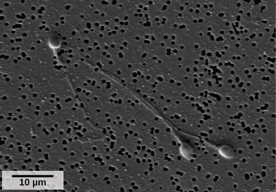
Semen is a mixture of sperm (about five percent of the total) and fluids from accessory glands that contribute most of the semen’s volume. Sperm are haploid cells, consisting of a flagellum for motility, a neck that contains the cell’s energy-producing mitochondria, and a head that contains the genetic material (Figure 18.11). An acrosome (acrosomal vesicle) is found at the top of the head of the sperm. This structure contains enzymes that can digest the protective coverings that surround the egg and allow the sperm to fuse with the egg. An ejaculate will contain from two to five milliliters of fluid and from 50–120 million sperm per milliliter.
Sperm form in the walls of seminiferous tubules that are coiled inside the testes (Figure 18.12; Table 18.1). The walls of the seminiferous tubules are made up of the developing sperm cells, with the least developed sperm at the periphery of the tubule and the fully developed sperm next to the lumen. The sperm cells are associated with Sertoli cells that nourish and promote the development of the sperm. Other cells present between the walls of the tubules are the interstitial cells of Leydig, which produce testosterone once the male reaches adolescence.
When the sperm have developed flagella they leave the seminiferous tubules and enter the epididymis (Figure 18.12; Table 18.1). This structure lies along the top and posterior of the testes and is the site of sperm maturation. The sperm leave the epididymis and enter the vas deferens, which carries the sperm behind the bladder, and forms the ejaculatory duct with the duct from the seminal vesicles. During a vasectomy, a section of the vas deferens is removed, preventing sperm (but not the secretions of the accessory glands) from being passed out of the body during ejaculation and preventing fertilization.
The bulk of the semen comes from the accessory glands associated with the male reproductive system. These are the seminal vesicles, the prostate gland, and the bulbourethral gland (Figure 18.12; Table 18.1). The secretions from the accessory glands provide important compounds for the sperm including nutrients, electrolytes, and pH buffering. There are also coagulation factors that affect sperm delivery and motility.
Which of the following statements about the male reproductive system is false?
- The vas deferens carries sperm from the testes to the seminal vesicles.
- The ejaculatory duct joins the urethra.
- Both the prostate and the bulbourethral glands produce components of the semen.
- The prostate gland is located in the testes.
| Male Reproductive Anatomy | ||
|---|---|---|
| Organ | Location | Function |
| Scrotum | External | Supports testes and regulates their temperature |
| Penis | External | Delivers urine, copulating organ |
| Testes | Internal | Produce sperm and male hormones |
| Seminal Vesicles | Internal | Contribute to semen production |
| Prostate Gland | Internal | Contributes to semen production |
| Bulbourethtral Glands | Internal | Neutralize urine in urethra |
Female Reproductive Anatomy
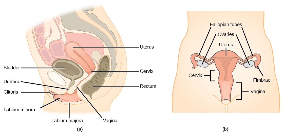
A number of female reproductive structures are exterior to the body. These include the breasts and the vulva, which consists of the mons pubis, clitoris, labia majora, labia minora, and the vestibular glands (Figure 18.13; Table 18.2).
The breasts consist of mammary glands and fat. Each gland consists of 15 to 25 lobes that have ducts that empty at the nipple and that supply the nursing child with nutrient- and antibody-rich milk to aid development and protect the child.
Internal female reproductive structures include ovaries, oviducts, the uterus, and the vagina (Figure 18.13; Table 18.2). The pair of ovaries is held in place in the abdominal cavity by a system of ligaments. The outermost layer of the ovary is made up of follicles, each consisting of one or more follicular cells that surround, nourish, and protect a single egg. During the menstrual period, a batch of follicular cells develops and prepares their eggs for release. At ovulation, one follicle ruptures and one egg is released. Following ovulation, the follicular tissue that surrounded the ovulated egg stays within the ovary and grows to form a solid mass called the corpus luteum. The corpus luteum secretes additional estrogen and the hormone progesterone that helps maintain the uterine lining during pregnancy. The ovaries also produce hormones, such as estrogen.
The oviducts, or fallopian tubes, extend from the uterus in the lower abdominal cavity to the ovaries, but they are not in contact with the ovaries. The lateral ends of the oviducts flare out into a trumpet-like structure and have a fringe of finger-like projections called fimbrae. When an egg is released at ovulation, the fimbrae help the nonmotile egg enter into the tube. The walls of the oviducts have a ciliated epithelium over smooth muscle. The cilia beat, and the smooth muscle contracts, moving the egg toward the uterus. Fertilization usually takes place within the oviduct and the developing embryo is moved toward the uterus. It usually takes the egg or embryo a week to travel through the oviduct.
Sterilization in women is called a tubal ligation; it is analogous to a vasectomy in males in that the oviducts are severed and sealed, preventing sperm from reaching the egg.
The uterus is a structure about the size of a woman’s fist. The uterus has a thick muscular wall and is lined with an endometrium rich in blood vessels and mucus glands that develop and thicken during the female cycle. Thickening of the endometrium prepares the uterus to receive the fertilized egg or zygote, which will then implant itself in the endometrium. The uterus supports the developing embryo and fetus during gestation. Contractions of the smooth muscle in the uterus aid in forcing the baby through the vagina during labor. If fertilization does not occur, a portion of the lining of the uterus sloughs off during each menstrual period. The endometrium builds up again in preparation for implantation. Part of the uterus, called the cervix, protrudes into the top of the vagina.
The vagina is a muscular tube that serves several purposes. It allows menstrual flow to leave the body. It is the receptacle for the penis during intercourse and the pathway for the delivery of offspring.
| Female Reproductive Anatomy | ||
|---|---|---|
| Organ | Location | Function |
| Clitoris | External | Sensory organ |
| Mons pubis | External | Fatty area overlying pubic bone |
| Labia majora | External | Covers labia minora; contains sweat and sebaceous glands |
| Labia minora | External | Covers vestibule |
| Greater vestibular glands | External | Secrete mucus; lubricate vagina |
| Breast | External | Produces and delivers milk |
| Ovaries | Internal | Produce and develop eggs |
| Oviducts | Internal | Transport egg to uterus; site of fertilization |
| Uterus | Internal | Supports developing embryo |
| Vagina | Internal | Common tube for intercourse, birth canal, passing menstrual flow |
Gametogenesis (Spermatogenesis and Oogenesis)
Gametogenesis, the production of sperm and eggs, involves the process of meiosis. During meiosis, two nuclear divisions separate the paired chromosomes in the nucleus and then separate the chromatids that were made during an earlier stage of the cell’s life cycle. Meiosis and its associated cell divisions produces haploid cells with half of each pair of chromosomes normally found in diploid cells. The production of sperm is called spermatogenesis and the production of eggs is called oogenesis.
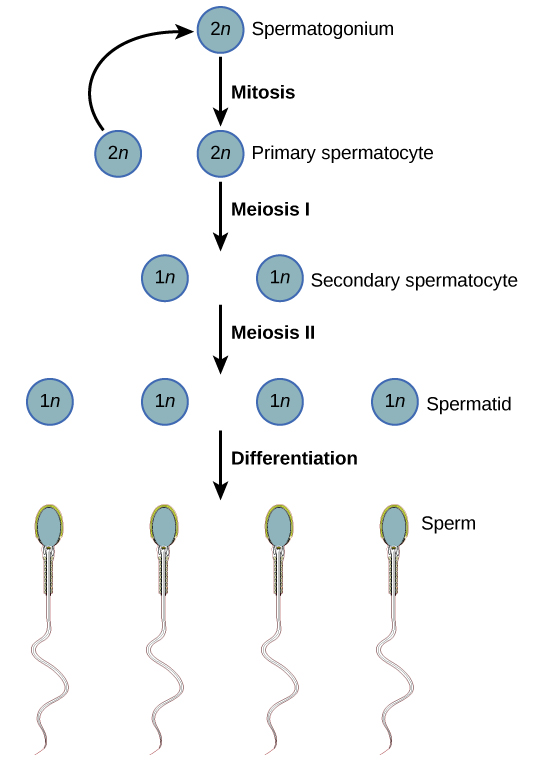
Spermatogenesis
Spermatogenesis occurs in the wall of the seminiferous tubules, with the most primitive cells at the periphery of the tube and the most mature sperm at the lumen of the tube (Figure 18.14). Immediately under the capsule of the tubule are diploid, undifferentiated cells. These stem cells, each called a spermatogonium (pl. spermatogonia), go through mitosis to produce one cell that remains as a stem cell and a second cell called a primary spermatocyte that will undergo meiosis to produce sperm.
The diploid primary spermatocyte goes through meiosis I to produce two haploid cells called secondary spermatocytes. Each secondary spermatocyte divides after meiosis II to produce two cells called spermatids. The spermatids eventually reach the lumen of the tubule and grow a flagellum, becoming sperm cells. Four sperm result from each primary spermatocyte that goes through meiosis.

Visit this site to see the process of spermatogenesis.
Oogenesis
Oogenesis occurs in the outermost layers of the ovaries. As with sperm production, oogenesis starts with a germ cell. In oogenesis, this germ cell is called an oogonium and forms during the embryological development of the individual. The oogonium undergoes mitosis to produce about one to two million oocytes by the time of birth.
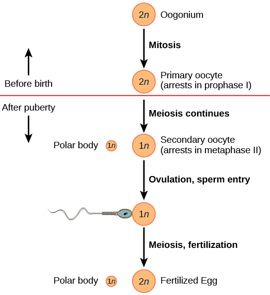
The primary oocytes begin meiosis before birth (Figure 18.15). However, the meiotic division is arrested in its progress in the first prophase stage. At the time of birth, all future eggs are in prophase I. This situation is in contrast with the male reproductive system in which sperm are produced continuously throughout the life of the individual. Starting at adolescence, anterior pituitary hormones cause the development of a few follicles in an ovary each month. This results in a primary oocyte finishing the first meiotic division. The cell divides unequally, with most of the cytoplasm and organelles going to one cell, called a secondary oocyte, and only one set of chromosomes and a small amount of cytoplasm going to the other cell. This second cell is called a polar body and usually dies. Cell division is again arrested, this time at metaphase II. At ovulation, this secondary oocyte is released and travels toward the uterus through the oviduct. If the secondary oocyte is fertilized, the cell continues through meiosis II, producing a second polar body and haploid egg, which fuses with the haploid sperm to form a fertilized egg (zygote) containing all 46 chromosomes.
Hormonal Control of Reproduction
The human male and female reproductive cycles are controlled by the interaction of hormones from the hypothalamus and anterior pituitary with hormones from reproductive tissues and organs. In both sexes, the hypothalamus monitors and causes the release of hormones from the anterior pituitary gland. When the reproductive hormone is required, the hypothalamus sends a gonadotropin-releasing hormone (GnRH) to the anterior pituitary. This causes the release of follicle stimulating hormone (FSH) and luteinizing hormone (LH) from the anterior pituitary into the blood. Although these hormones are named after their functions in female reproduction, they are produced in both sexes and play important roles in controlling reproduction. Other hormones have specific functions in the male and female reproductive systems.
Male Hormones
At the onset of puberty, the hypothalamus causes the release of FSH and LH into the male system for the first time. FSH enters the testes and stimulates the Sertoli cells located in the walls of the seminiferous tubules to begin promoting spermatogenesis (Figure 18.16). LH also enters the testes and stimulates the interstitial cells of Leydig, located in between the walls of the seminiferous tubules, to make and release testosterone into the testes and the blood.
Testosterone stimulates spermatogenesis. This hormone is also responsible for the secondary sexual characteristics that develop in the male during adolescence. The secondary sex characteristics in males include a deepening of the voice, the growth of facial, axillary, and pubic hair, an increase in muscle bulk, and the beginnings of the sex drive.
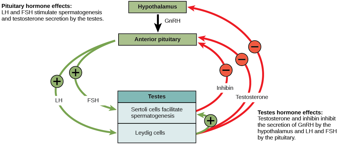
A negative feedback system occurs in the male with rising levels of testosterone acting on the hypothalamus and anterior pituitary to inhibit the release of GnRH, FSH, and LH. In addition, the Sertoli cells produce the hormone inhibin, which is released into the blood when the sperm count is too high. This inhibits the release of GnRH and FSH, which will cause spermatogenesis to slow down. If the sperm count reaches a low of 20 million/mL, the Sertoli cells cease the release of inhibin, and the sperm count increases.
Female Hormones
The control of reproduction in females is more complex. The female reproductive cycle is divided into the ovarian cycle and the menstrual cycle. The ovarian cycle governs the preparation of endocrine tissues and release of eggs, while the menstrual cycle governs the preparation and maintenance of the uterine lining (Figure 18.17). These cycles are coordinated over a 22–32 day cycle, with an average length of 28 days.
As with the male, the GnRH from the hypothalamus causes the release of the hormones FSH and LH from the anterior pituitary. In addition, estrogen and progesterone are released from the developing follicles. As with testosterone in males, estrogen is responsible for the secondary sexual characteristics of females. These include breast development, flaring of the hips, and a shorter period for bone growth.
The Ovarian Cycle and the Menstrual Cycle
The ovarian and menstrual cycles are regulated by hormones of the hypothalamus, pituitary, and ovaries (Figure 18.17). The ebb and flow of the hormones causes the ovarian and menstrual cycles to advance. The ovarian and menstrual cycles occur concurrently. The first half of the ovarian cycle is the follicular phase. Slowly rising levels of FSH cause the growth of follicles on the surface of the ovary. This process prepares the egg for ovulation. As the follicles grow, they begin releasing estrogen. The first few days of this cycle coincide with menstruation or the sloughing off of the functional layer of the endometrium in the uterus. After about five days, estrogen levels rise and the menstrual cycle enters the proliferative phase. The endometrium begins to regrow, replacing the blood vessels and glands that deteriorated during the end of the last cycle.
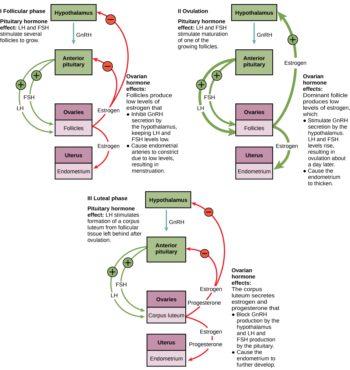
Which of the following statements about hormone regulation of the female reproductive cycle is false?
- LH and FSH are produced in the pituitary, and estrogen and progesterone are produced in the ovaries.
- Estradiol and progesterone secreted from the corpus luteum cause the endometrium to thicken.
- Both progesterone and estrogen are produced by the follicles.
- Secretion of GnRH by the hypothalamus is inhibited by low levels of estrogen but stimulated by high levels of estrogen.
Just prior to the middle of the cycle (approximately day 14), the high level of estrogen causes FSH and especially LH to rise rapidly then fall. The spike in LH causes the most mature follicle to rupture and release its egg. This is ovulation. The follicles that did not rupture degenerate and their eggs are lost. The level of estrogen decreases when the extra follicles degenerate.
Following ovulation, the ovarian cycle enters its luteal phase and the menstrual cycle enters its secretory phase, both of which run from about day 15 to 28. The luteal and secretory phases refer to changes in the ruptured follicle. The cells in the follicle undergo physical changes and produce a structure called a corpus luteum. The corpus luteum produces estrogen and progesterone. The progesterone facilitates the regrowth of the uterine lining and inhibits the release of further FSH and LH. The uterus is being prepared to accept a fertilized egg, should it occur during this cycle. The inhibition of FSH and LH prevents any further eggs and follicles from developing, while the progesterone is elevated. The level of estrogen produced by the corpus luteum increases to a steady level for the next few days.
If no fertilized egg is implanted into the uterus, the corpus luteum degenerates and the levels of estrogen and progesterone decrease. The endometrium begins to degenerate as the progesterone levels drop, initiating the next menstrual cycle. The decrease in progesterone also allows the hypothalamus to send GnRH to the anterior pituitary, releasing FSH and LH and starting the cycles again.
A reproductive endocrinologist is a physician who treats a variety of hormonal disorders related to reproduction and infertility in both men and women. The disorders include menstrual problems, infertility, pregnancy loss, sexual dysfunction, and menopause. Doctors may use fertility drugs, surgery, or assisted reproductive techniques (ART) in their therapy. ART involves the use of procedures to manipulate the egg or sperm to facilitate reproduction, such as in vitro fertilization.
Reproductive endocrinologists undergo extensive medical training, first in a four-year residency in obstetrics and gynecology, then in a three-year fellowship in reproductive endocrinology. To be board certified in this area, the physician must pass written and oral exams in both areas.
Gestation
Pregnancy begins with the fertilization of an egg and continues through to the birth of the individual. The length of time of gestation, or the gestation period, in humans is 266 days and is similar in other great apes.
Within 24 hours of fertilization, the egg nucleus has finished meiosis and the egg and sperm nuclei fuse. With fusion, the cell is known as a zygote. The zygote initiates cleavage and the developing embryo travels through the oviduct to the uterus. The developing embryo must implant into the wall of the uterus within seven days, or it will deteriorate and die. The outer layers of the developing embryo or blastocyst grow into the endometrium by digesting the endometrial cells, and healing of the endometrium closes up the blastocyst into the tissue. Another layer of the blastocyst, the chorion, begins releasing a hormone called human beta chorionic gonadotropin (β-HCG), which makes its way to the corpus luteum and keeps that structure active. This ensures adequate levels of progesterone that will maintain the endometrium of the uterus for the support of the developing embryo. Pregnancy tests determine the level of β-HCG in urine or serum. If the hormone is present, the test is positive.
The gestation period is divided into three equal periods or trimesters. During the first two-to-four weeks of the first trimester, nutrition and waste are handled by the endometrial lining through diffusion. As the trimester progresses, the outer layer of the embryo begins to merge with the endometrium, and the placenta forms. The placenta takes over the nutrient and waste requirements of the embryo and fetus, with the mother’s blood passing nutrients to the placenta and removing waste from it. Chemicals from the fetus, such as bilirubin, are processed by the mother’s liver for elimination. Some of the mother’s immunoglobulins will pass through the placenta, providing passive immunity against some potential infections.
Internal organs and body structures begin to develop during the first trimester. By five weeks, limb buds, eyes, the heart, and liver have been basically formed. By eight weeks, the term fetus applies, and the body is essentially formed (Figure 18.18a). The individual is about five centimeters (two inches) in length and many of the organs, such as the lungs and liver, are not yet functioning. Exposure to any toxins is especially dangerous during the first trimester, as all of the body’s organs and structures are going through initial development. Anything that interferes with chemical signaling during that development can have a severe effect on the fetus’ survival.
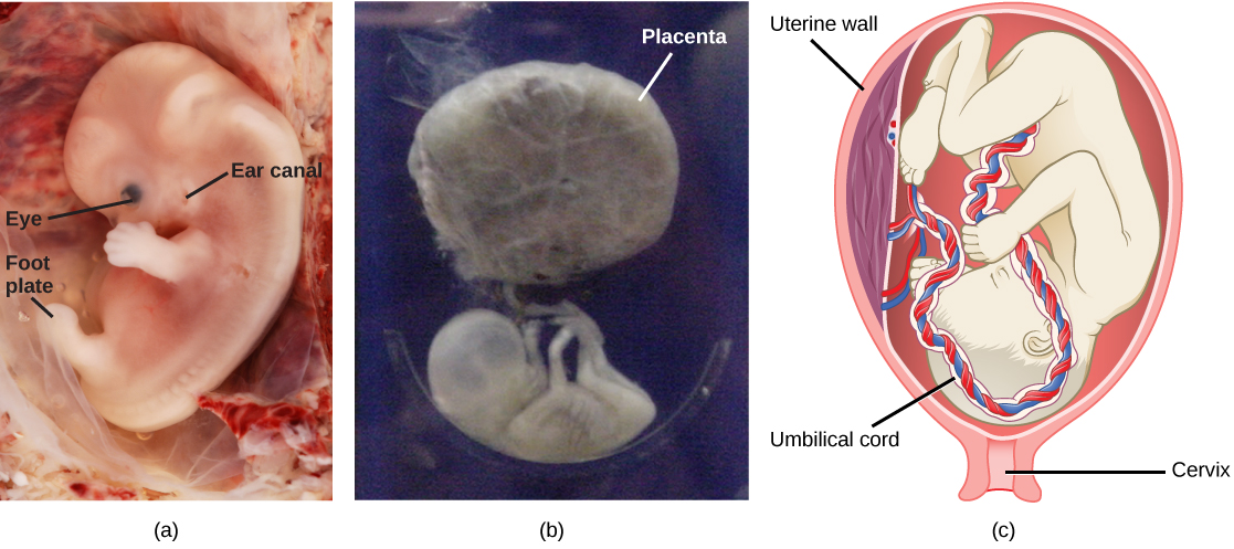
During the second trimester, the fetus grows to about 30 cm (about 12 inches) (Figure 18.18b). It becomes active and the mother usually feels the first movements. All organs and structures continue to develop. The placenta has taken over the functions of nutrition and waste elimination and the production of estrogen and progesterone from the corpus luteum, which has degenerated. The placenta will continue functioning up through the delivery of the baby. During the third trimester, the fetus grows to 3 to 4 kg (6.5–8.5 lbs.) and about 50 cm (19–20 inches) long (Figure 18.18c). This is the period of the most rapid growth during the pregnancy as all organ systems continue to grow and develop.

Visit this website to see the stages of human fetal development.
Labor is the muscular contractions to expel the fetus and placenta from the uterus. Toward the end of the third trimester, estrogen causes receptors on the uterine wall to develop and bind the hormone oxytocin. At this time, the baby reorients, facing forward and down with the back or crown of the head engaging the cervix (uterine opening). This causes the cervix to stretch and nerve impulses are sent to the hypothalamus, which signals the release of oxytocin from the posterior pituitary. Oxytocin causes smooth muscle in the uterine wall to contract. At the same time, the placenta releases prostaglandins into the uterus, increasing the contractions. A positive feedback relay occurs between the uterus, hypothalamus, and the posterior pituitary to assure an adequate supply of oxytocin. As more smooth muscle cells are recruited, the contractions increase in intensity and force.
There are three stages to labor. During stage one, the cervix thins and dilates. This is necessary for the baby and placenta to be expelled during birth. The cervix will eventually dilate to about 10 cm. During stage two, the baby is expelled from the uterus. The uterus contracts and the mother pushes as she compresses her abdominal muscles to aid the delivery. The last stage is the passage of the placenta after the baby has been born and the organ has completely disengaged from the uterine wall. If labor should stop before stage two is reached, synthetic oxytocin, known as Pitocin, can be administered to restart and maintain labor.
Glossary
- asexual reproduction
- a mechanism that produces offspring that are genetically identical to the parent
- blastocyst
- the structure formed when cells in the mammalian blastula separate into an inner and outer layer
- budding
- a form of asexual reproduction that results from the outgrowth of a part of an organism leading to a separation from the original animal into two individuals
- bulbourethral gland
- the paired glands in the human male that produce a secretion that cleanses the urethra prior to ejaculation
- clitoris
- a sensory and erectile structure in female mammals, homologous to the male penis, stimulated during sexual arousal
- corpus luteum
- the endocrine tissue that develops from an ovarian follicle after ovulation; secretes progesterone and estrogen during pregnancy
- estrogen
- a reproductive hormone in females that assists in endometrial regrowth, ovulation, and calcium absorption
- external fertilization
- the fertilization of eggs by sperm outside an animal’s body, often during spawning
- fission
- (also, binary fission) a form of asexual reproduction in which an organism splits into two separate organisms or two parts that regenerate the missing portions of the body
- follicle stimulating hormone (FSH)
- a reproductive hormone that causes sperm production in men and follicle development in women
- fragmentation
- the breaking of an organism into parts and the growth of a separate individual from each part
- gastrulation
- the process in which the blastula folds over itself to form the three germ layers
- gestation period
- the length of time of development, from conception to birth, of the young of a viviparous animal
- gestation
- the development before birth of a viviparous animal
- gonadotropin-releasing hormone (GnRH)
- a hormone from the hypothalamus that causes the release of FSH and LH from the anterior pituitary
- hermaphroditism
- the state of having both male and female reproductive structures within the same individual
- human beta chorionic gonadotropin (β-HCG)
- a hormone produced by the chorion of the zygote that helps to maintain the corpus luteum and elevated levels of progesterone
- inhibin
- a hormone made by Sertoli cells, provides negative feedback to hypothalamus in control of FSH and GnRH release
- inner cell mass
- the inner layer of cells in the blastocyst, which becomes the embryo
- internal fertilization
- the fertilization of eggs by sperm inside the body of the female
- interstitial cell of Leydig
- a cell type found next to the seminiferous tubules that makes testosterone
- labia majora
- the large folds of tissue covering inguinal area
- labia minora
- the smaller folds of tissue within labia majora
- luteinizing hormone (LH)
- a reproductive hormone in both men and women, causes testosterone production in men and ovulation and lactation in women
- menstrual cycle
- the cycle of the degradation and re-growth of the endometrium
- oogenesis
- the process of producing haploid eggs
- organogenesis
- the process of organ formation during development
- ovarian cycle
- the cycle of preparation of egg for ovulation and the conversion of the follicle to the corpus luteum
- oviduct
- (also, fallopian tube) the muscular tube connecting uterus with ovary area
- oviparity
- a process by which fertilized eggs are laid outside the female’s body and develop there, receiving nourishment from the yolk that is a part of the egg
- ovoviparity
- a process by which fertilized eggs are retained within the female; the embryo obtains its nourishment from the egg’s yolk, and the young are fully developed when they are hatched
- ovulation
- the release of an oocyte from a mature follicle in the ovary of a vertebrate
- parthenogenesis
- a form of asexual reproduction in which an egg develops into a complete individual without being fertilized
- penis
- the male reproductive structure for urine elimination and copulation
- placenta
- the organ that supports the transport of nutrients and waste between the mothers and fetus’ blood in eutherian mammals
- progesterone
- a reproductive hormone in women; assists in endometrial regrowth and inhibition of FSH and LH release
- prostate gland
- a structure that is a mixture of smooth muscle and glandular material and that contributes to semen
- Sertoli cell
- a cell in the walls of the seminiferous tubules that assists developing sperm and secretes inhibin
- scrotum
- a sac containing testes, exterior to body
- semen
- a fluid mixture of sperm and supporting materials
- seminal vesicle
- a secretory accessory gland in male; contributes to semen
- seminiferous tubule
- the structures within which sperm production occurs in the testes
- sex determination
- the mechanism by which the sex of individuals in sexually reproducing organisms is initially established
- sexual reproduction
- a form of reproduction in which cells containing genetic material from two individuals combines to produce genetically unique offspring
- spermatogenesis
- the process of producing haploid sperm
- testes
- a pair of male reproductive organs
- testosterone
- a reproductive hormone in men that assists in sperm production and promoting secondary sexual characteristics
- trophoblast
- the outer layer of cells in the blastocyst, which gives rise to the embryo’s contribution to the placenta
- uterus
- a female reproductive structure in which an embryo develops
- vagina
- a muscular tube for the passage of menstrual flow, copulation, and birth of offspring
- viviparity
- a process in which the young develop within the female and are born in a nonembryonic state
- zona pellucida
- the protective layer around the mammalian egg
<!–CNX: Start Area: “Sections Summary”–>
Reproduction may be asexual when one individual produces genetically identical offspring, or sexual when the genetic material from two individuals is combined to produce genetically diverse offspring. Asexual reproduction in animals occurs through fission, budding, fragmentation, and parthenogenesis. Sexual reproduction may involve fertilization inside the body or in the external environment. A species may have separate sexes or combined sexes; when the sexes are combined they may be expressed at different times in the life cycle. The sex of an individual may be determined by various chromosomal systems or environmental factors such as temperature.
Sexual reproduction starts with the combination of a sperm and an egg in a process called fertilization. This can occur either outside the bodies or inside the female. The method of fertilization varies among animals. Some species release the egg and sperm into the environment, some species retain the egg and receive the sperm into the female body and then expel the developing embryo covered with shell, while still other species retain the developing offspring throughout the gestation period.
The early stages of embryonic development begin with fertilization. The process of fertilization is tightly controlled to ensure that only one sperm fuses with one egg. After fertilization, the zygote undergoes cleavage to form the blastula. The blastula, which in some species is a hollow ball of cells, undergoes a process called gastrulation, during which the three germ layers form. The ectoderm gives rise to the nervous system and the epidermal skin cells, the mesoderm gives rise to the muscle cells and connective tissue in the body, and the endoderm gives rise to the digestive system and other internal organs. Organogenesis is the formation of organs from the germ layers. Each germ layer gives rise to specific tissue types.
The reproductive structures that evolved in land animals allow males and females to mate, fertilize internally, and support the growth and development of offspring. Gametogenesis, the production of sperm (spermatogenesis) and eggs (oogenesis), takes place through the process of meiosis.
The male and female reproductive cycles are controlled by hormones released from the hypothalamus and anterior pituitary and hormones from reproductive tissues and organs. The hypothalamus monitors the need for FSH and LH production and release from the anterior pituitary. FSH and LH affect reproductive structures to cause the formation of sperm and the preparation of eggs for release and possible fertilization. In the male, FSH and LH stimulate Sertoli cells and interstitial cells of Leydig in the testes to facilitate sperm production. The Leydig cells produce testosterone, which also is responsible for the secondary sexual characteristics of males. In females, FSH and LH cause estrogen and progesterone to be produced. They regulate the female reproductive cycle, which is divided into the ovarian cycle and the menstrual cycle.
Human pregnancy begins with fertilization of an egg and proceeds through the three trimesters of gestation. The first trimester lays down the basic structures of the body, including the limb buds, heart, eyes, and the liver. The second trimester continues the development of all of the organs and systems. The third trimester exhibits the greatest growth of the fetus and culminates in labor and delivery. The labor process has three stages (contractions, delivery of the fetus, and expulsion of the placenta), each propelled by hormones.
<!–CNX: Start Area: “Art Connections”–>
Figure 18.12 Which of the following statements about the male reproductive system is false?
- The vas deferens carries sperm from the testes to the seminal vesicles.
- The ejaculatory duct joins the urethra.
- Both the prostate and the bulbourethral glands produce components of the semen.
- The prostate gland is located in the testes.
- LH and FSH are produced in the pituitary, and estrogen and progesterone are produced in the ovaries.
- Estradiol and progesterone secreted from the corpus luteum cause the endometrium to thicken.
- Both progesterone and estrogen are produced by the follicles.
- Secretion of GnRH by the hypothalamus is inhibited by low levels of estrogen but stimulated by high levels of estrogen.
<!–CNX: Start Area: “Multiple Choice”–>
- chickens
- bees
- rabbits
- sea stars
B
- sexual reproduction
- parthenogenesis
- budding
- fragmentation
A
- aquatic
- forested
- savanna
- steppe
A
- blastula
- zygote
- organs
- germ layers
D
- ectoderm
- endoderm
- mesoderm
- none of the above
A
- scrotum
- seminal vesicles
- seminiferous tubules
- prostate gland
C
- labia minora
- breast
- ovaries
- uterus
D
Which hormone causes FSH and LH to be released?
- testosterone
- estrogen
- GnRH
- progesterone
C
- the placenta
- diffusion through the endometrium
- the chorion
- the blastocyst
B
- oxytocin
- estrogen
- β-HCG
- progesterone
A
<!–CNX: Start Area: “Free Response”–>
Temperatures can vary from year to year and an unusually cold or hot year might produce offspring all of one sex, making it hard for individuals to find mates.
A possible advantage of hermaphroditism might be that anytime an individual of the same species is encountered a mating is possible, unlike separate sexes that must find an individual of the right sex to mate. (Also, every individual in a hermaphrodite population is able to produce offspring, which is not the case in populations with separate sexes.) A disadvantage might be that hermaphrodite populations are less efficient because they do not specialize in one sex or another, which means a hermaphrodite does not produce as many offspring through eggs or sperm as do species with separate sexes. (Other answers are possible.)
What do you think would happen if multiple sperm fused with one egg?
If multiple sperm fused with one egg, a zygote with a multiple ploidy level (multiple copies of the chromosomes) would form, and then would die.
Stem cells are laid down in the male during gestation and lie dormant until adolescence. Stem cells in the female increase to one to two million and enter the first meiotic division and are arrested in prophase. At adolescence, spermatogenesis begins and continues until death, producing the maximum number of sperm with each meiotic division. Oogenesis continues again at adolescence in batches of eggs with each menstrual cycle. These primary oocytes finish the first meiotic division, producing a viable egg with most of the cytoplasm and its contents, and a second cell called a polar body containing 23 chromosomes. The second meiotic division is initiated and arrested in metaphase. At ovulation, one egg is released. If this egg is fertilized, it finishes the second meiotic division. This is a diploid, fertilized egg.
Low levels of progesterone allow the hypothalamus to send GnRH to the anterior pituitary and cause the release of FSH and LH. FSH stimulates follicles on the ovary to grow and prepare the eggs for ovulation. As the follicles increase in size, they begin to release estrogen and a low level of progesterone into the blood. The level of estrogen rises to a peak, causing a spike in the concentration of LH. This causes the most mature follicle to rupture and ovulation occurs.
Stage one of labor results in uterine contractions, which thin the cervix and dilate the cervical opening. Stage two delivers the baby, and stage three delivers the placenta.
<!–CNX: Start Area: “Art Connections”–>
<!–CNX: Start Area: “Multiple Choice”–>
B
A
A
D
A
C
D
C
B
A
<!–CNX: Start Area: “Free Response”–>
Temperatures can vary from year to year and an unusually cold or hot year might produce offspring all of one sex, making it hard for individuals to find mates.
A possible advantage of hermaphroditism might be that anytime an individual of the same species is encountered a mating is possible, unlike separate sexes that must find an individual of the right sex to mate. (Also, every individual in a hermaphrodite population is able to produce offspring, which is not the case in populations with separate sexes.) A disadvantage might be that hermaphrodite populations are less efficient because they do not specialize in one sex or another, which means a hermaphrodite does not produce as many offspring through eggs or sperm as do species with separate sexes. (Other answers are possible.)
If multiple sperm fused with one egg, a zygote with a multiple ploidy level (multiple copies of the chromosomes) would form, and then would die.
Stem cells are laid down in the male during gestation and lie dormant until adolescence. Stem cells in the female increase to one to two million and enter the first meiotic division and are arrested in prophase. At adolescence, spermatogenesis begins and continues until death, producing the maximum number of sperm with each meiotic division. Oogenesis continues again at adolescence in batches of eggs with each menstrual cycle. These primary oocytes finish the first meiotic division, producing a viable egg with most of the cytoplasm and its contents, and a second cell called a polar body containing 23 chromosomes. The second meiotic division is initiated and arrested in metaphase. At ovulation, one egg is released. If this egg is fertilized, it finishes the second meiotic division. This is a diploid, fertilized egg.
Low levels of progesterone allow the hypothalamus to send GnRH to the anterior pituitary and cause the release of FSH and LH. FSH stimulates follicles on the ovary to grow and prepare the eggs for ovulation. As the follicles increase in size, they begin to release estrogen and a low level of progesterone into the blood. The level of estrogen rises to a peak, causing a spike in the concentration of LH. This causes the most mature follicle to rupture and ovulation occurs.
Stage one of labor results in uterine contractions, which thin the cervix and dilate the cervical opening. Stage two delivers the baby, and stage three delivers the placenta.