OpenStax College, OpenStax College
Chapter 16. The Body’s Systems

Introduction*
The arctic fox, a complex animal that has adapted to its environment, illustrates the relationships between an animal’s form and function. The multicellular bodies of animals consist of tissues that make up more complex organs and organ systems. The organ systems of an animal maintain homeostasis within the multicellular body. These systems are adapted to obtain the necessary nutrients and other resources needed by the cells of the body, to remove the wastes those cells produce, to coordinate the activities of the cells, tissues, and organs throughout the body, and to coordinate the many responses of the individual organism to its environment.
16.1. Homeostasis and Osmoregulation*
By the end of this section, you will be able to:
- Explain the concept of homeostasis
- Describe thermoregulation of endothermic and ectothermic animals
- Explain how the kidneys serve as the main osmoregulatory organs in the human body
Homeostasis refers to the relatively stable state inside the body of an animal. Animal organs and organ systems constantly adjust to internal and external changes in order to maintain this steady state. Examples of internal conditions maintained homeostatically are the level of blood glucose, body temperature, blood calcium level. These conditions remain stable because of physiologic processes that result in negative feedback relationships. If the blood glucose or calcium rises, this sends a signal to organs responsible for lowering blood glucose or calcium. The signals that restore the normal levels are examples of negative feedback. When homeostatic mechanisms fail, the results can be unfavorable for the animal. Homeostatic mechanisms keep the body in dynamic equilibrium by constantly adjusting to the changes that the body’s systems encounter. Even an animal that is apparently inactive is maintaining this homeostatic equilibrium. Two examples of factors that are regulated homeostatically are temperature and water content. The processes that maintain homeostasis of these two factors are called thermoregulation and osmoregulation.
Homeostasis
The goal of homeostasis is the maintenance of equilibrium around a specific value of some aspect of the body or its cells called a set point. While there are normal fluctuations from the set point, the body’s systems will usually attempt to go back to this point. A change in the internal or external environment is called a stimulus and is detected by a receptor; the response of the system is to adjust the activities of the system so the value moves back toward the set point. For instance, if the body becomes too warm, adjustments are made to cool the animal. If glucose levels in the blood rise after a meal, adjustments are made to lower them and to get the nutrient into tissues that need it or to store it for later use.
When a change occurs in an animal’s environment, an adjustment must be made so that the internal environment of the body and cells remains stable. The receptor that senses the change in the environment is part of a feedback mechanism. The stimulus—temperature, glucose, or calcium levels—is detected by the receptor. The receptor sends information to a control center, often the brain, which relays appropriate signals to an effector organ that is able to cause an appropriate change, either up or down, depending on the information the sensor was sending.
Thermoregulation
Animals can be divided into two groups: those that maintain a constant body temperature in the face of differing environmental temperatures, and those that have a body temperature that is the same as their environment and thus varies with the environmental temperature. Animals that do not have internal control of their body temperature are called ectotherms. The body temperature of these organisms is generally similar to the temperature of the environment, although the individual organisms may do things that keep their bodies slightly below or above the environmental temperature. This can include burrowing underground on a hot day or resting in the sunlight on a cold day. The ectotherms have been called cold-blooded, a term that may not apply to an animal in the desert with a very warm body temperature.
An animal that maintains a constant body temperature in the face of environmental changes is called an endotherm. These animals are able to maintain a level of activity that an ectothermic animal cannot because they generate internal heat that keeps their cellular processes operating optimally even when the environment is cold.

Watch this Discovery Channel video on thermoregulation to see illustrations of the process in a variety of animals.
Animals conserve or dissipate heat in a variety of ways. Endothermic animals have some form of insulation. They have fur, fat, or feathers. Animals with thick fur or feathers create an insulating layer of air between their skin and internal organs. Polar bears and seals live and swim in a subfreezing environment and yet maintain a constant, warm, body temperature. The arctic fox, for example, uses its fluffy tail as extra insulation when it curls up to sleep in cold weather. Mammals can increase body heat production by shivering, which is an involuntary increase in muscle activity. In addition, arrector pili muscles can contract causing individual hairs to stand up when the individual is cold. This increases the insulating effect of the hair. Humans retain this reaction, which does not have the intended effect on our relatively hairless bodies; it causes “goose bumps” instead. Mammals use layers of fat as insulation also. Loss of significant amounts of body fat will compromise an individual’s ability to conserve heat.
Ectotherms and endotherms use their circulatory systems to help maintain body temperature. Vasodilation, the opening up of arteries to the skin by relaxation of their smooth muscles, brings more blood and heat to the body surface, facilitating radiation and evaporative heat loss, cooling the body. Vasoconstriction, the narrowing of blood vessels to the skin by contraction of their smooth muscles, reduces blood flow in peripheral blood vessels, forcing blood toward the core and vital organs, conserving heat. Some animals have adaptions to their circulatory system that enable them to transfer heat from arteries to veins that are flowing next to each other, warming blood returning to the heart. This is called a countercurrent heat exchange; it prevents the cold venous blood from cooling the heart and other internal organs. The countercurrent adaptation is found in dolphins, sharks, bony fish, bees, and hummingbirds.
Some ectothermic animals use changes in their behavior to help regulate body temperature. They simply seek cooler areas during the hottest part of the day in the desert to keep from getting too warm. The same animals may climb onto rocks in the evening to capture heat on a cold desert night before entering their burrows.
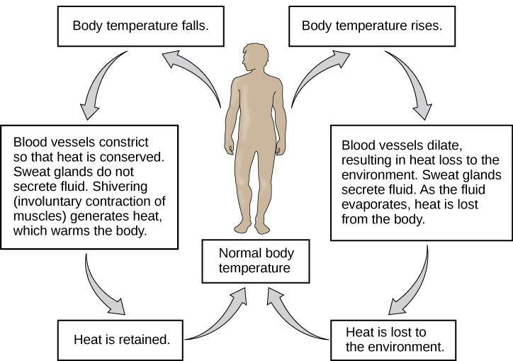
Thermoregulation is coordinated by the nervous system (Figure 16.2). The processes of temperature control are centered in the hypothalamus of the advanced animal brain. The hypothalamus maintains the set point for body temperature through reflexes that cause vasodilation or vasoconstriction and shivering or sweating. The sympathetic nervous system under control of the hypothalamus directs the responses that effect the changes in temperature loss or gain that return the body to the set point. The set point may be adjusted in some instances. During an infection, compounds called pyrogens are produced and circulate to the hypothalamus resetting the thermostat to a higher value. This allows the body’s temperature to increase to a new homeostatic equilibrium point in what is commonly called a fever. The increase in body heat makes the body less optimal for bacterial growth and increases the activities of cells so they are better able to fight the infection.
When bacteria are destroyed by leukocytes, pyrogens are released into the blood. Pyrogens reset the body’s thermostat to a higher temperature, resulting in fever. How might pyrogens cause the body temperature to rise?
Osmoregulation
Osmoregulation is the process of maintaining salt and water balance ( osmotic balance) across membranes within the body. The fluids inside and surrounding cells are composed of water, electrolytes, and nonelectrolytes. An electrolyte is a compound that dissociates into ions when dissolved in water. A nonelectrolyte, in contrast, does not dissociate into ions in water. The body’s fluids include blood plasma, fluid that exists within cells, and the interstitial fluid that exists in the spaces between cells and tissues of the body. The membranes of the body (both the membranes around cells and the “membranes” made of cells lining body cavities) are semipermeable membranes. Semipermeable membranes are permeable to certain types of solutes and to water, but typically cell membranes are impermeable to solutes.
The body does not exist in isolation. There is a constant input of water and electrolytes into the system. Excess water, electrolytes, and wastes are transported to the kidneys and excreted, helping to maintain osmotic balance. Insufficient fluid intake results in fluid conservation by the kidneys. Biological systems constantly interact and exchange water and nutrients with the environment by way of consumption of food and water and through excretion in the form of sweat, urine, and feces. Without a mechanism to regulate osmotic pressure, or when a disease damages this mechanism, there is a tendency to accumulate toxic waste and water, which can have dire consequences.
Mammalian systems have evolved to regulate not only the overall osmotic pressure across membranes, but also specific concentrations of important electrolytes in the three major fluid compartments: blood plasma, interstitial fluid, and intracellular fluid. Since osmotic pressure is regulated by the movement of water across membranes, the volume of the fluid compartments can also change temporarily. Since blood plasma is one of the fluid components, osmotic pressures have a direct bearing on blood pressure.
Excretory System
The human excretory system functions to remove waste from the body through the skin as sweat, the lungs in the form of exhaled carbon dioxide, and through the urinary system in the form of urine. All three of these systems participate in osmoregulation and waste removal. Here we focus on the urinary system, which is comprised of the paired kidneys, the ureter, urinary bladder and urethra (Figure 16.3). The kidneys are a pair of bean-shaped structures that are located just below the liver in the body cavity. Each of the kidneys contains more than a million tiny units called nephrons that filter blood containing the metabolic wastes from cells. All the blood in the human body is filtered about 60 times a day by the kidneys. The nephrons remove wastes, concentrate them, and form urine that is collected in the bladder.
Internally, the kidney has three regions—an outer cortex, a medulla in the middle, and the renal pelvis, which is the expanded end of the ureter. The renal cortex contains the nephrons—the functional unit of the kidney. The renal pelvis collects the urine and leads to the ureter on the outside of the kidney. The ureters are urine-bearing tubes that exit the kidney and empty into the urinary bladder.
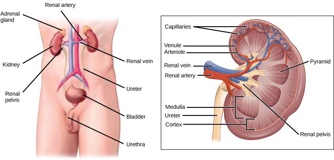
Blood enters each kidney from the aorta, the main artery supplying the body below the heart, through a renal artery. It is distributed in smaller vessels until it reaches each nephron in capillaries. Within the nephron the blood comes in intimate contact with the waste-collecting tubules in a structure called the glomerulus. Water and many solutes present in the blood, including ions of sodium, calcium, magnesium, and others; as well as wastes and valuable substances such as amino acids, glucose and vitamins, leave the blood and enter the tubule system of the nephron. As materials pass through the tubule much of the water, required ions, and useful compounds are reabsorbed back into the capillaries that surround the tubules leaving the wastes behind. Some of this reabsorption requires active transport and consumes ATP. Some wastes, including ions and some drugs remaining in the blood, diffuse out of the capillaries into the interstitial fluid and are taken up by the tubule cells. These wastes are then actively secreted into the tubules. The blood then collects in larger and larger vessels and leaves the kidney in the renal vein. The renal vein joins the inferior vena cava, the main vein that returns blood to the heart from the lower body. The amounts of water and ions reabsorbed into the circulatory system are carefully regulated and this is an important way the body regulates its water content and ion levels. The waste is collected in larger tubules and then leaves the kidney in the ureter, which leads to the bladder where urine, the combination of waste materials and water, is stored.
The bladder contains sensory nerves, stretch receptors that signal when it needs to be emptied. These signals create the urge to urinate, which can be voluntarily suppressed up to a limit. The conscious decision to urinate sets in play signals that open the sphincters, rings of smooth muscle that close off the opening, to the urethra that allows urine to flow out of the bladder and the body.
Dialysis is a medical process of removing wastes and excess water from the blood by diffusion and ultrafiltration. When kidney function fails, dialysis must be done to artificially rid the body of wastes and fluids. This is a vital process to keep patients alive. In some cases, the patients undergo artificial dialysis until they are eligible for a kidney transplant. In others who are not candidates for kidney transplants, dialysis is a lifelong necessity.
Dialysis technicians typically work in hospitals and clinics. While some roles in this field include equipment development and maintenance, most dialysis technicians work in direct patient care. Their on-the-job duties, which typically occur under the direct supervision of a registered nurse, focus on providing dialysis treatments. This can include reviewing patient history and current condition, assessing and responding to patient needs before and during treatment, and monitoring the dialysis process. Treatment may include taking and reporting a patient’s vital signs, preparing solutions and equipment to ensure accurate and sterile procedures.
16.2. Digestive System*
By the end of this section, you will be able to:
- Explain the processes of digestion and absorption
- Explain the specialized functions of the organs involved in processing food in the body
- Describe the ways in which organs work together to digest food and absorb nutrients
- Describe the essential nutrients required for cellular function that cannot be synthesized by the animal body
- Describe how excess carbohydrates and energy are stored in the body
All living organisms need nutrients to survive. While plants can obtain nutrients from their roots and the energy molecules required for cellular function through the process of photosynthesis, animals obtain their nutrients by the consumption of other organisms. At the cellular level, the biological molecules necessary for animal function are amino acids, lipid molecules, nucleotides, and simple sugars. However, the food consumed consists of protein, fat, and complex carbohydrates. Animals must convert these macromolecules into the simple molecules required for maintaining cellular function. The conversion of the food consumed to the nutrients required is a multistep process involving digestion and absorption. During digestion, food particles are broken down to smaller components, which are later absorbed by the body. This happens by both physical means, such as chewing, and by chemical means.
One of the challenges in human nutrition is maintaining a balance between food intake, storage, and energy expenditure. Taking in more food energy than is used in activity leads to storage of the excess in the form of fat deposits. The rise in obesity and the resulting diseases like type 2 diabetes makes understanding the role of diet and nutrition in maintaining good health all the more important.
The Human Digestive System
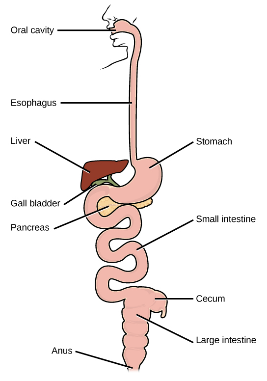
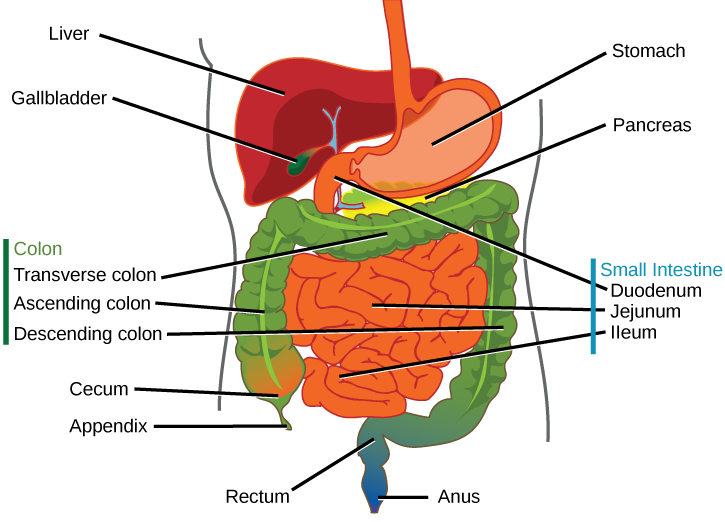
The process of digestion begins in the mouth with the intake of food (Figure 16.4). The teeth play an important role in masticating (chewing) or physically breaking food into smaller particles. The enzymes present in saliva also begin to chemically break down food. The food is then swallowed and enters the esophagus—a long tube that connects the mouth to the stomach. Using peristalsis, or wave-like smooth-muscle contractions, the muscles of the esophagus push the food toward the stomach. The stomach contents are extremely acidic, with a pH between 1.5 and 2.5. This acidity kills microorganisms, breaks down food tissues, and activates digestive enzymes. Further breakdown of food takes place in the small intestine where bile produced by the liver, and enzymes produced by the small intestine and the pancreas, continue the process of digestion. The smaller molecules are absorbed into the blood stream through the epithelial cells lining the walls of the small intestine. The waste material travels on to the large intestine where water is absorbed and the drier waste material is compacted into feces; it is stored until it is excreted through the anus.
Oral Cavity
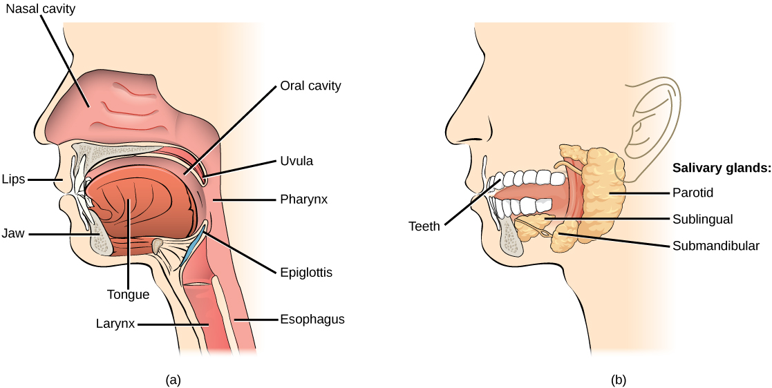
Both physical and chemical digestion begin in the mouth or oral cavity, which is the point of entry of food into the digestive system. The food is broken into smaller particles by mastication, the chewing action of the teeth. All mammals have teeth and can chew their food to begin the process of physically breaking it down into smaller particles.
The chemical process of digestion begins during chewing as food mixes with saliva, produced by the salivary glands (Figure 16.5). Saliva contains mucus that moistens food and buffers the pH of the food. Saliva also contains lysozyme, which has antibacterial action. It also contains an enzyme called salivary amylase that begins the process of converting starches in the food into a disaccharide called maltose. Another enzyme called lipase is produced by cells in the tongue to break down fats. The chewing and wetting action provided by the teeth and saliva prepare the food into a mass called the bolus for swallowing. The tongue helps in swallowing—moving the bolus from the mouth into the pharynx. The pharynx opens to two passageways: the esophagus and the trachea. The esophagus leads to the stomach and the trachea leads to the lungs. The epiglottis is a flap of tissue that covers the tracheal opening during swallowing to prevent food from entering the lungs.
Esophagus
The esophagus is a tubular organ that connects the mouth to the stomach. The chewed and softened food passes through the esophagus after being swallowed. The smooth muscles of the esophagus undergo peristalsis that pushes the food toward the stomach. The peristaltic wave is unidirectional—it moves food from the mouth the stomach, and reverse movement is not possible, except in the case of the vomit reflex. The peristaltic movement of the esophagus is an involuntary reflex; it takes place in response to the act of swallowing.
Ring-like muscles called sphincters form valves in the digestive system. The gastro-esophageal sphincter (or cardiac sphincter) is located at the stomach end of the esophagus. In response to swallowing and the pressure exerted by the bolus of food, this sphincter opens, and the bolus enters the stomach. When there is no swallowing action, this sphincter is shut and prevents the contents of the stomach from traveling up the esophagus. Acid reflux or “heartburn” occurs when the acidic digestive juices escape into the esophagus.
Stomach
A large part of protein digestion occurs in the stomach (Figure 16.7). The stomach is a saclike organ that secretes gastric digestive juices.
Protein digestion is carried out by an enzyme called pepsin in the stomach chamber. The highly acidic environment kills many microorganisms in the food and, combined with the action of the enzyme pepsin, results in the catabolism of protein in the food. Chemical digestion is facilitated by the churning action of the stomach caused by contraction and relaxation of smooth muscles. The partially digested food and gastric juice mixture is called chyme. Gastric emptying occurs within two to six hours after a meal. Only a small amount of chyme is released into the small intestine at a time. The movement of chyme from the stomach into the small intestine is regulated by hormones, stomach distension and muscular reflexes that influence the pyloric sphincter.
The stomach lining is unaffected by pepsin and the acidity because pepsin is released in an inactive form and the stomach has a thick mucus lining that protects the underlying tissue.
Small Intestine
Chyme moves from the stomach to the small intestine. The small intestine is the organ where the digestion of protein, fats, and carbohydrates is completed. The small intestine is a long tube-like organ with a highly folded surface containing finger-like projections called the villi. The top surface of each villus has many microscopic projections called microvilli. The epithelial cells of these structures absorb nutrients from the digested food and release them to the bloodstream on the other side. The villi and microvilli, with their many folds, increase the surface area of the small intestine and increase absorption efficiency of the nutrients.
The human small intestine is over 6 m (19.6 ft) long and is divided into three parts: the duodenum, the jejunum and the ileum. The duodenum is separated from the stomach by the pyloric sphincter. The chyme is mixed with pancreatic juices, an alkaline solution rich in bicarbonate that neutralizes the acidity of chyme from the stomach. Pancreatic juices contain several digestive enzymes that break down starches, disaccharides, proteins, and fats. Bile is produced in the liver and stored and concentrated in the gallbladder; it enters the duodenum through the bile duct. Bile contains bile salts, which make lipids accessible to the water-soluble enzymes. The monosaccharides, amino acids, bile salts, vitamins, and other nutrients are absorbed by the cells of the intestinal lining.
The undigested food is sent to the colon from the ileum via peristaltic movements. The ileum ends and the large intestine begins at the ileocecal valve. The vermiform, “worm-like,” appendix is located at the ileocecal valve. The appendix of humans has a minor role in immunity.
Large Intestine
The large intestine reabsorbs the water from indigestible food material and processes the waste material (Figure 16.6). The human large intestine is much smaller in length compared to the small intestine but larger in diameter. It has three parts: the cecum, the colon, and the rectum. The cecum joins the ileum to the colon and is the receiving pouch for the waste matter. The colon is home to many bacteria or “intestinal flora” that aid in the digestive processes. The colon has four regions, the ascending colon, the transverse colon, the descending colon and the sigmoid colon. The main functions of the colon are to extract the water and mineral salts from undigested food, and to store waste material.
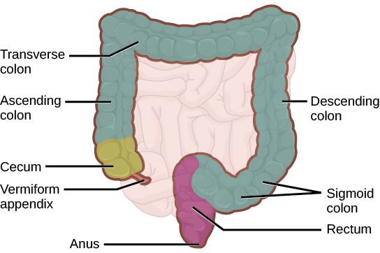
The rectum (Figure 16.6) stores feces until defecation. The feces are propelled using peristaltic movements during elimination. The anus is an opening at the far-end of the digestive tract and is the exit point for the waste material. Two sphincters regulate the exit of feces, the inner sphincter is involuntary and the outer sphincter is voluntary.
Accessory Organs
The organs discussed above are the organs of the digestive tract through which food passes. Accessory organs add secretions and enzymes that break down food into nutrients. Accessory organs include the salivary glands, the liver, the pancreas, and the gall bladder. The secretions of the liver, pancreas, and gallbladder are regulated by hormones in response to food consumption.
The liver is the largest internal organ in humans and it plays an important role in digestion of fats and detoxifying blood. The liver produces bile, a digestive juice that is required for the breakdown of fats in the duodenum. The liver also processes the absorbed vitamins and fatty acids and synthesizes many plasma proteins. The gallbladder is a small organ that aids the liver by storing bile and concentrating bile salts.
The pancreas secretes bicarbonate that neutralizes the acidic chyme and a variety of enzymes for the digestion of protein and carbohydrates.
Which of the following statements about the digestive system is false?
- Chyme is a mixture of food and digestive juices that is produced in the stomach.
- Food enters the large intestine before the small intestine.
- In the small intestine, chyme mixes with bile, which emulsifies fats.
- The stomach is separated from the small intestine by the pyloric sphincter.
Nutrition
The human diet should be well balanced to provide nutrients required for bodily function and the minerals and vitamins required for maintaining structure and regulation necessary for good health and reproductive capability (Figure 16.8).


Explore this interactive United States Department of Agriculture website to learn more about each food group and the recommended daily amounts.
The organic molecules required for building cellular material and tissues must come from food. During digestion, digestible carbohydrates are ultimately broken down into glucose and used to provide energy within the cells of the body. Complex carbohydrates, including polysaccharides, can be broken down into glucose through biochemical modification; however, humans do not produce the enzyme necessary to digest cellulose (fiber). The intestinal flora in the human gut are able to extract some nutrition from these plant fibers. These plant fibers are known as dietary fiber and are an important component of the diet. The excess sugars in the body are converted into glycogen and stored for later use in the liver and muscle tissue. Glycogen stores are used to fuel prolonged exertions, such as long-distance running, and to provide energy during food shortage. Fats are stored under the skin of mammals for insulation and energy reserves.
Proteins in food are broken down during digestion and the resulting amino acids are absorbed. All of the proteins in the body must be formed from these amino-acid constituents; no proteins are obtained directly from food.
Fats add flavor to food and promote a sense of satiety or fullness. Fatty foods are also significant sources of energy, and fatty acids are required for the construction of lipid membranes. Fats are also required in the diet to aid the absorption of fat-soluble vitamins and the production of fat-soluble hormones.
While the animal body can synthesize many of the molecules required for function from precursors, there are some nutrients that must be obtained from food. These nutrients are termed essential nutrients, meaning they must be eaten, because the body cannot produce them.
The fatty acids omega-3 alpha-linolenic acid and omega-6 linoleic acid are essential fatty acids needed to make some membrane phospholipids. Vitamins are another class of essential organic molecules that are required in small quantities. Many of these assist enzymes in their function and, for this reason, are called coenzymes. Absence or low levels of vitamins can have a dramatic effect on health. Minerals are another set of inorganic essential nutrients that must be obtained from food. Minerals perform many functions, from muscle and nerve function, to acting as enzyme cofactors. Certain amino acids also must be procured from food and cannot be synthesized by the body. These amino acids are the “essential” amino acids. The human body can synthesize only 11 of the 20 required amino acids; the rest must be obtained from food.
With obesity at high rates in the United States, there is a public health focus on reducing obesity and associated health risks, which include diabetes, colon and breast cancer, and cardiovascular disease. How does the food consumed contribute to obesity?
Fatty foods are calorie-dense, meaning that they have more calories per unit mass than carbohydrates or proteins. One gram of carbohydrates has four calories, one gram of protein has four calories, and one gram of fat has nine calories. Animals tend to seek lipid-rich food for their higher energy content. Greater amounts of food energy taken in than the body’s requirements will result in storage of the excess in fat deposits.
Excess carbohydrate is used by the liver to synthesize glycogen. When glycogen stores are full, additional glucose is converted into fatty acids. These fatty acids are stored in adipose tissue cells—the fat cells in the mammalian body whose primary role is to store fat for later use.
The rate of obesity among children is rapidly rising in the United States. To combat childhood obesity and ensure that children get a healthy start in life, in 2010 First Lady Michelle Obama launched the Let’s Move! campaign. The goal of this campaign is to educate parents and caregivers on providing healthy nutrition and encouraging active lifestyles in future generations. This program aims to involve the entire community, including parents, teachers, and healthcare providers to ensure that children have access to healthy foods—more fruits, vegetables, and whole grains—and consume fewer calories from processed foods. Another goal is to ensure that children get physical activity. With the increase in television viewing and stationary pursuits such as video games, sedentary lifestyles have become the norm. Visit www.letsmove.gov to learn more.
16.3. Circulatory and Respiratory Systems*
By the end of this section, you will be able to:
- Describe the passage of air from the outside environment to the lungs
- Describe the function of the circulatory system
- Describe the cardiac cycle
- Explain how blood flows through the body
Animals are complex multicellular organisms that require a mechanism for transporting nutrients throughout their bodies and removing wastes. The human circulatory system has a complex network of blood vessels that reach all parts of the body. This extensive network supplies the cells, tissues, and organs with oxygen and nutrients, and removes carbon dioxide and waste compounds.
The medium for transport of gases and other molecules is the blood, which continually circulates through the system. Pressure differences within the system cause the movement of the blood and are created by the pumping of the heart.
Gas exchange between tissues and the blood is an essential function of the circulatory system. In humans, other mammals, and birds, blood absorbs oxygen and releases carbon dioxide in the lungs. Thus the circulatory and respiratory system, whose function is to obtain oxygen and discharge carbon dioxide, work in tandem.
The Respiratory System
Take a breath in and hold it. Wait several seconds and then let it out. Humans, when they are not exerting themselves, breathe approximately 15 times per minute on average. This equates to about 900 breaths an hour or 21,600 breaths per day. With every inhalation, air fills the lungs, and with every exhalation, it rushes back out. That air is doing more than just inflating and deflating the lungs in the chest cavity. The air contains oxygen that crosses the lung tissue, enters the bloodstream, and travels to organs and tissues. There, oxygen is exchanged for carbon dioxide, which is a cellular waste material. Carbon dioxide exits the cells, enters the bloodstream, travels back to the lungs, and is expired out of the body during exhalation.
Breathing is both a voluntary and an involuntary event. How often a breath is taken and how much air is inhaled or exhaled is regulated by the respiratory center in the brain in response to signals it receives about the carbon dioxide content of the blood. However, it is possible to override this automatic regulation for activities such as speaking, singing and swimming under water.
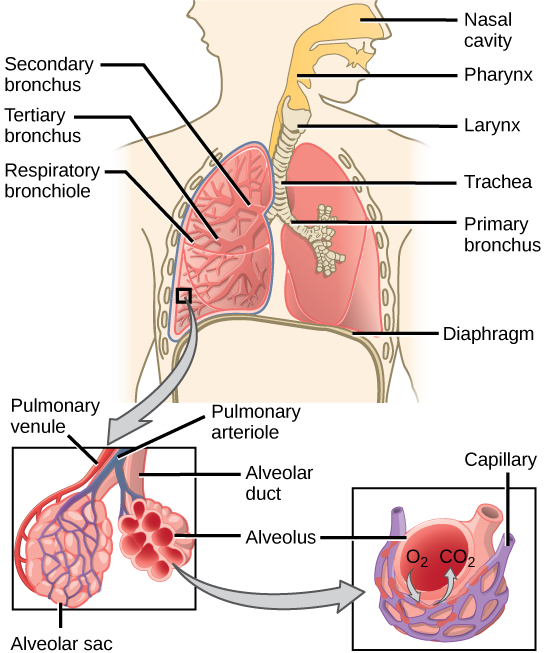
During inhalation the diaphragm descends creating a negative pressure around the lungs and they begin to inflate, drawing in air from outside the body. The air enters the body through the nasal cavity located just inside the nose (Figure 16.9). As the air passes through the nasal cavity, the air is warmed to body temperature and humidified by moisture from mucous membranes. These processes help equilibrate the air to the body conditions, reducing any damage that cold, dry air can cause. Particulate matter that is floating in the air is removed in the nasal passages by hairs, mucus, and cilia. Air is also chemically sampled by the sense of smell.
From the nasal cavity, air passes through the pharynx (throat) and the larynx (voice box) as it makes its way to the trachea (Figure 16.9). The main function of the trachea is to funnel the inhaled air to the lungs and the exhaled air back out of the body. The human trachea is a cylinder, about 25 to 30 cm (9.8–11.8 in) long, which sits in front of the esophagus and extends from the pharynx into the chest cavity to the lungs. It is made of incomplete rings of cartilage and smooth muscle. The cartilage provides strength and support to the trachea to keep the passage open. The trachea is lined with cells that have cilia and secrete mucus. The mucus catches particles that have been inhaled, and the cilia move the particles toward the pharynx.
The end of the trachea divides into two bronchi that enter the right and left lung. Air enters the lungs through the primary bronchi. The primary bronchus divides, creating smaller and smaller diameter bronchi until the passages are under 1 mm (.03 in) in diameter when they are called bronchioles as they split and spread through the lung. Like the trachea, the bronchus and bronchioles are made of cartilage and smooth muscle. Bronchi are innervated by nerves of both the parasympathetic and sympathetic nervous systems that control muscle contraction (parasympathetic) or relaxation (sympathetic) in the bronchi and bronchioles, depending on the nervous system’s cues. The final bronchioles are the respiratory bronchioles. Alveolar ducts are attached to the end of each respiratory bronchiole. At the end of each duct are alveolar sacs, each containing 20 to 30 alveoli. Gas exchange occurs only in the alveoli. The alveoli are thin-walled and look like tiny bubbles within the sacs. The alveoli are in direct contact with capillaries of the circulatory system. Such intimate contact ensures that oxygen will diffuse from the alveoli into the blood. In addition, carbon dioxide will diffuse from the blood into the alveoli to be exhaled. The anatomical arrangement of capillaries and alveoli emphasizes the structural and functional relationship of the respiratory and circulatory systems. Estimates for the surface area of alveoli in the lungs vary around 100 m2. This large area is about the area of half a tennis court. This large surface area, combined with the thin-walled nature of the alveolar cells, allows gases to easily diffuse across the cells.
Which of the following statements about the human respiratory system is false?
- When we breathe in, air travels from the pharynx to the trachea.
- The bronchioles branch into bronchi.
- Alveolar ducts connect to alveolar sacs.
- Gas exchange between the lungs and blood takes place in the alveolus.

Watch this video for a review of the respiratory system.
The Circulatory System
The circulatory system is a network of vessels—the arteries, veins, and capillaries—and a pump, the heart. In all vertebrate organisms this is a closed-loop system, in which the blood is largely separated from the body’s other extracellular fluid compartment, the interstitial fluid, which is the fluid bathing the cells. Blood circulates inside blood vessels and circulates unidirectionally from the heart around one of two circulatory routes, then returns to the heart again; this is a closed circulatory system. Open circulatory systems are found in invertebrate animals in which the circulatory fluid bathes the internal organs directly even though it may be moved about with a pumping heart.
The Heart
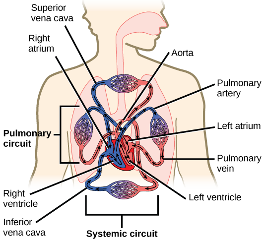
The heart is a complex muscle that consists of two pumps: one that pumps blood through pulmonary circulation to the lungs, and the other that pumps blood through systemic circulation to the rest of the body’s tissues (and the heart itself).
The heart is asymmetrical, with the left side being larger than the right side, correlating with the different sizes of the pulmonary and systemic circuits (Figure 16.10). In humans, the heart is about the size of a clenched fist; it is divided into four chambers: two atria and two ventricles. There is one atrium and one ventricle on the right side and one atrium and one ventricle on the left side. The right atrium receives deoxygenated blood from the systemic circulation through the major veins: the superior vena cava, which drains blood from the head and from the veins that come from the arms, as well as the inferior vena cava, which drains blood from the veins that come from the lower organs and the legs. This deoxygenated blood then passes to the right ventricle through the tricuspid valve, which prevents the backflow of blood. After it is filled, the right ventricle contracts, pumping the blood to the lungs for reoxygenation. The left atrium receives the oxygen-rich blood from the lungs. This blood passes through the bicuspid valve to the left ventricle where the blood is pumped into the aorta. The aorta is the major artery of the body, taking oxygenated blood to the organs and muscles of the body. This pattern of pumping is referred to as double circulation and is found in all mammals. (Figure 16.10).
Which of the following statements about the circulatory system is false?
- Blood in the pulmonary vein is deoxygenated.
- Blood in the inferior vena cava is deoxygenated.
- Blood in the pulmonary artery is deoxygenated.
- Blood in the aorta is oxygenated.
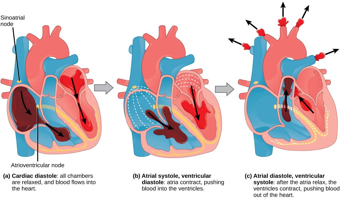
The Cardiac Cycle
The main purpose of the heart is to pump blood through the body; it does so in a repeating sequence called the cardiac cycle. The cardiac cycle is the flow of blood through the heart coordinated by electrochemical signals that cause the heart muscle to contract and relax. In each cardiac cycle, a sequence of contractions pushes out the blood, pumping it through the body; this is followed by a relaxation phase, where the heart fills with blood. These two phases are called the systole (contraction) and diastole (relaxation), respectively (Figure 16.11). The signal for contraction begins at a location on the outside of the right atrium. The electrochemical signal moves from there across the atria causing them to contract. The contraction of the atria forces blood through the valves into the ventricles. Closing of these valves caused by the contraction of the ventricles produces a “lub” sound. The signal has, by this time, passed down the walls of the heart, through a point between the right atrium and right ventricle. The signal then causes the ventricles to contract. The ventricles contract together forcing blood into the aorta and the pulmonary arteries. Closing of the valves to these arteries caused by blood being drawn back toward the heart during ventricular relaxation produces a monosyllabic “dub” sound.
The pumping of the heart is a function of the cardiac muscle cells, or cardiomyocytes, that make up the heart muscle. Cardiomyocytes are distinctive muscle cells that are striated like skeletal muscle but pump rhythmically and involuntarily like smooth muscle; adjacent cells are connected by intercalated disks found only in cardiac muscle. These connections allow the electrical signal to travel directly to neighboring muscle cells.
The electrical impulses in the heart produce electrical currents that flow through the body and can be measured on the skin using electrodes. This information can be observed as an electrocardiogram (ECG) a recording of the electrical impulses of the cardiac muscle.

Visit the following website to see the heart’s pacemaker, or electrocardiogram system, in action.
Blood Vessels
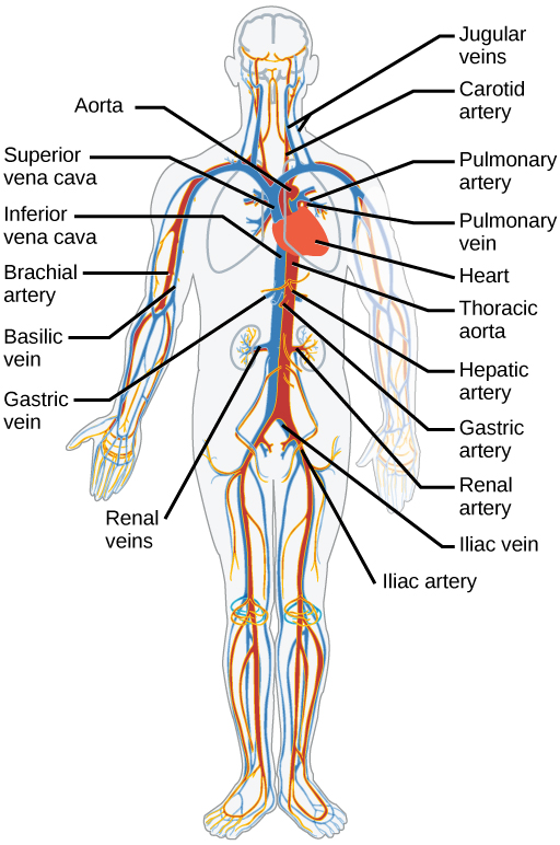
The blood from the heart is carried through the body by a complex network of blood vessels (Figure 16.12). Arteries take blood away from the heart. The main artery of the systemic circulation is the aorta; it branches into major arteries that take blood to different limbs and organs. The aorta and arteries near the heart have heavy but elastic walls that respond to and smooth out the pressure differences caused by the beating heart. Arteries farther away from the heart have more muscle tissue in their walls that can constrict to affect flow rates of blood. The major arteries diverge into minor arteries, and then smaller vessels called arterioles, to reach more deeply into the muscles and organs of the body.
Arterioles diverge into capillary beds. Capillary beds contain a large number, 10’s to 100’s of capillaries that branch among the cells of the body. Capillaries are narrow-diameter tubes that can fit single red blood cells and are the sites for the exchange of nutrients, waste, and oxygen with tissues at the cellular level. Fluid also leaks from the blood into the interstitial space from the capillaries. The capillaries converge again into venules that connect to minor veins that finally connect to major veins. Veins are blood vessels that bring blood high in carbon dioxide back to the heart. Veins are not as thick-walled as arteries, since pressure is lower, and they have valves along their length that prevent backflow of blood away from the heart. The major veins drain blood from the same organs and limbs that the major arteries supply.
16.4. Endocrine System*
By the end of this section, you will be able to:
- List the different types of hormones and explain their roles in maintaining homeostasis
- Explain how hormones work
- Explain how hormone production is regulated
- Describe the role of different glands in the endocrine system
- Explain how the different glands work together to maintain homeostasis
The endocrine system produces hormones that function to control and regulate many different body processes. The endocrine system coordinates with the nervous system to control the functions of the other organ systems. Cells of the endocrine system produce molecular signals called hormones. These cells may compose endocrine glands, may be tissues or may be located in organs or tissues that have functions in addition to hormone production. Hormones circulate throughout the body and stimulate a response in cells that have receptors able to bind with them. The changes brought about in the receiving cells affect the functioning of the organ system to which they belong. Many of the hormones are secreted in response to signals from the nervous system, thus the two systems act in concert to effect changes in the body.
Hormones
Maintaining homeostasis within the body requires the coordination of many different systems and organs. One mechanism of communication between neighboring cells, and between cells and tissues in distant parts of the body, occurs through the release of chemicals called hormones. Hormones are released into body fluids, usually blood, which carries them to their target cells where they elicit a response. The cells that secrete hormones are often located in specific organs, called endocrine glands, and the cells, tissues, and organs that secrete hormones make up the endocrine system. Examples of endocrine organs include the pancreas, which produces the hormones insulin and glucagon to regulate blood-glucose levels, the adrenal glands, which produce hormones such as epinephrine and norepinephrine that regulate responses to stress, and the thyroid gland, which produces thyroid hormones that regulate metabolic rates.
The endocrine glands differ from the exocrine glands. Exocrine glands secrete chemicals through ducts that lead outside the gland (not to the blood). For example, sweat produced by sweat glands is released into ducts that carry sweat to the surface of the skin. The pancreas has both endocrine and exocrine functions because besides releasing hormones into the blood. It also produces digestive juices, which are carried by ducts into the small intestine.
An endocrinologist is a medical doctor who specializes in treating endocrine disorders. An endocrine surgeon specializes in the surgical treatment of endocrine diseases and glands. Some of the diseases that are managed by endocrinologists include disorders of the pancreas (diabetes mellitus), disorders of the pituitary (gigantism, acromegaly, and pituitary dwarfism), disorders of the thyroid gland (goiter and Graves’ disease), and disorders of the adrenal glands (Cushing’s disease and Addison’s disease).
Endocrinologists are required to assess patients and diagnose endocrine disorders through extensive use of laboratory tests. Many endocrine diseases are diagnosed using tests that stimulate or suppress endocrine organ functioning. Blood samples are then drawn to determine the effect of stimulating or suppressing an endocrine organ on the production of hormones. For example, to diagnose diabetes mellitus, patients are required to fast for 12 to 24 hours. They are then given a sugary drink, which stimulates the pancreas to produce insulin to decrease blood-glucose levels. A blood sample is taken one to two hours after the sugar drink is consumed. If the pancreas is functioning properly, the blood-glucose level will be within a normal range. Another example is the A1C test, which can be performed during blood screening. The A1C test measures average blood-glucose levels over the past two to three months. The A1C test is an indicator of how well blood glucose is being managed over a long time.
Once a disease such as diabetes has been diagnosed, endocrinologists can prescribe lifestyle changes and medications to treat the disease. Some cases of diabetes mellitus can be managed by exercise, weight loss, and a healthy diet; in other cases, medications may be required to enhance insulin’s production or effect. If the disease cannot be controlled by these means, the endocrinologist may prescribe insulin injections.
In addition to clinical practice, endocrinologists may also be involved in primary research and development activities. For example, ongoing islet transplant research is investigating how healthy pancreas islet cells may be transplanted into diabetic patients. Successful islet transplants may allow patients to stop taking insulin injections.
How Hormones Work
Hormones cause changes in target cells by binding to specific cell-surface or intracellular hormone receptors, molecules embedded in the cell membrane or floating in the cytoplasm with a binding site that matches a binding site on the hormone molecule. In this way, even though hormones circulate throughout the body and come into contact with many different cell types, they only affect cells that possess the necessary receptors. Receptors for a specific hormone may be found on or in many different cells or may be limited to a small number of specialized cells. For example, thyroid hormones act on many different tissue types, stimulating metabolic activity throughout the body. Cells can have many receptors for the same hormone but often also possess receptors for different types of hormones. The number of receptors that respond to a hormone determines the cell’s sensitivity to that hormone, and the resulting cellular response. Additionally, the number of receptors available to respond to a hormone can change over time, resulting in increased or decreased cell sensitivity. In up-regulation, the number of receptors increases in response to rising hormone levels, making the cell more sensitive to the hormone and allowing for more cellular activity. When the number of receptors decreases in response to rising hormone levels, called down-regulation, cellular activity is reduced.
Endocrine Glands
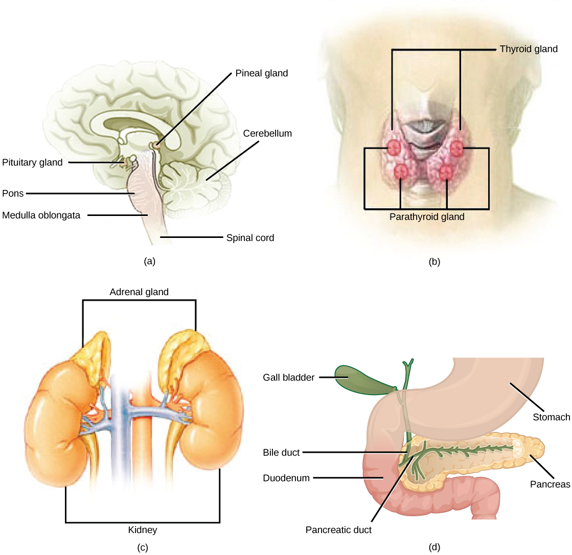
The endocrine glands secrete hormones into the surrounding interstitial fluid; those hormones then diffuse into blood and are carried to various organs and tissues within the body. The endocrine glands include the pituitary, thyroid, parathyroid, adrenal glands, gonads, pineal, and pancreas.
The pituitary gland, sometimes called the hypophysis, is located at the base of the brain (Figure 16.13a). It is attached to the hypothalamus. The posterior lobe stores and releases oxytocin and antidiuretic hormone produced by the hypothalamus. The anterior lobe responds to hormones produced by the hypothalamus by producing its own hormones, most of which regulate other hormone-producing glands.
The anterior pituitary produces six hormones: growth hormone, prolactin, thyroid-stimulating hormone, adrenocorticotropic hormone, follicle-stimulating hormone, and luteinizing hormone. Growth hormone stimulates cellular activities like protein synthesis that promote growth. Prolactin stimulates the production of milk by the mammary glands. The other hormones produced by the anterior pituitary regulate the production of hormones by other endocrine tissues (Table 16.1). The posterior pituitary is significantly different in structure from the anterior pituitary. It is a part of the brain, extending down from the hypothalamus, and contains mostly nerve fibers that extend from the hypothalamus to the posterior pituitary.
The thyroid gland is located in the neck, just below the larynx and in front of the trachea (Figure 16.13b). It is a butterfly-shaped gland with two lobes that are connected. The thyroid follicle cells synthesize the hormone thyroxine, which is also known as T4 because it contains four atoms of iodine, and triiodothyronine, also known as T3 because it contains three atoms of iodine. T3 and T4 are released by the thyroid in response tothyroid-stimulating hormone produced by the anterior pituitary, and both T3 and T4 have the effect of stimulating metabolic activity in the body and increasing energy use. A third hormone, calcitonin, is also produced by the thyroid. Calcitonin is released in response to rising calcium ion concentrations in the blood and has the effect of reducing those levels.
Most people have four parathyroid glands; however, the number can vary from two to six. These glands are located on the posterior surface of the thyroid gland (Figure 16.13b).
The parathyroid glands produce parathyroid hormone. Parathyroid hormone increases blood calcium concentrations when calcium ion levels fall below normal.
The adrenal glands are located on top of each kidney (Figure 16.13c). The adrenal glands consist of an outer adrenal cortex and an inner adrenal medulla. These regions secrete different hormones.
The adrenal cortex produces mineralocorticoids, glucocorticoids, and androgens. The main mineralocorticoid is aldosterone, which regulates the concentration of ions in urine, sweat, and saliva. Aldosterone release from the adrenal cortex is stimulated by a decrease in blood concentrations of sodium ions, blood volume, or blood pressure, or by an increase in blood potassium levels. The glucocorticoids maintain proper blood-glucose levels between meals. They also control a response to stress by increasing glucose synthesis from fats and proteins and interact with epinephrine to cause vasoconstriction. Androgens are sex hormones that are produced in small amounts by the adrenal cortex. They do not normally affect sexual characteristics and may supplement sex hormones released from the gonads. The adrenal medulla contains two types of secretory cells: one that produces epinephrine (adrenaline) and another that produces norepinephrine (noradrenaline). Epinephrine and norepinephrine cause immediate, short-term changes in response to stressors, inducing the so-called fight-or-flight response. The responses include increased heart rate, breathing rate, cardiac muscle contractions, and blood-glucose levels. They also accelerate the breakdown of glucose in skeletal muscles and stored fats in adipose tissue, and redirect blood flow toward skeletal muscles and away from skin and viscera. The release of epinephrine and norepinephrine is stimulated by neural impulses from the sympathetic nervous system that originate from the hypothalamus.
The pancreas is an elongate organ located between the stomach and the proximal portion of the small intestine (Figure 16.13d). It contains both exocrine cells that excrete digestive enzymes and endocrine cells that release hormones.
The endocrine cells of the pancreas form clusters called pancreatic islets or the islets of Langerhans. Among the cell types in each pancreatic islet are the alpha cells, which produce the hormone glucagon, and the beta cells, which produce the hormone insulin. These hormones regulate blood-glucose levels. Alpha cells release glucagon as blood-glucose levels decline. When blood-glucose levels rise, beta cells release insulin. Glucagon causes the release of glucose to the blood from the liver, and insulin facilitates the uptake of glucose by the body’s cells.
The gonads—the male testes and female ovaries—produce steroid hormones. The testes produce androgens, testosterone being the most prominent, which allow for the development of secondary sex characteristics and the production of sperm cells. The ovaries produce estrogen and progesterone, which cause secondary sex characteristics, regulate production of eggs, control pregnancy, and prepare the body for childbirth.
There are several organs whose primary functions are non-endocrine but that also possess endocrine functions. These include the heart, kidneys, intestines, thymus, and adipose tissue. The heart has endocrine cells in the walls of the atria that release a hormone in response to increased blood volume. It causes a reduction in blood volume and blood pressure, and reduces the concentration of Na+ in the blood.
The gastrointestinal tract produces several hormones that aid in digestion. The endocrine cells are located in the mucosa of the GI tract throughout the stomach and small intestine. They trigger the release of gastric juices, which help to break down and digest food in the GI tract.
The kidneys also possess endocrine function. Two of these hormones regulate ion concentrations and blood volume or pressure. Erythropoietin (EPO) is released by kidneys in response to low oxygen levels. EPO triggers the formation of red blood cells in the bone marrow. EPO has been used by athletes to improve performance. But EPO doping has its risks, since it thickens the blood and increases strain on the heart; it also increases the risk of blood clots and therefore heart attacks and stroke.
The thymus is found behind the sternum. The thymus produces hormones referred to as thymosins, which contribute to the development of the immune response in infants. Adipose tissue, or fat tissue, produces the hormone leptin in response to food intake. Leptin produces a feeling of satiety after eating, reducing the urge for further eating.
| Endocrine Glands and Their Associated Hormones | ||
|---|---|---|
| Endocrine Gland | Associated Hormones | Effect |
| Pituitary (anterior) | growth hormone | promotes growth of body tissues |
| prolactin | promotes milk production | |
| thyroid-stimulating hormone | stimulates thyroid hormone release | |
| adrenocorticotropic hormone | stimulates hormone release by adrenal cortex | |
| follicle-stimulating hormone | stimulates gamete production | |
| luteinizing hormone | stimulates androgen production by gonads in males; stimulates ovulation and production of estrogen and progesterone in females | |
| Pituitary (posterior) | antidiuretic hormone | stimulates water reabsorption by kidneys |
| oxytocin | stimulates uterine contractions during childbirth | |
| Thyroid | thyroxine, triiodothyronine | stimulate metabolism |
| calcitonin | reduces blood Ca2+ levels | |
| Parathyroid | parathyroid hormone | increases blood Ca2+ levels |
| Adrenal (cortex) | aldosterone | increases blood Na+ levels |
| cortisol, corticosterone, cortisone | increase blood-glucose levels | |
| Adrenal (medulla) | epinephrine, norepinephrine | stimulate fight-or-flight response |
| Pancreas | insulin | reduces blood-glucose levels |
| glucagon | increases blood-glucose levels | |
Regulation of Hormone Production
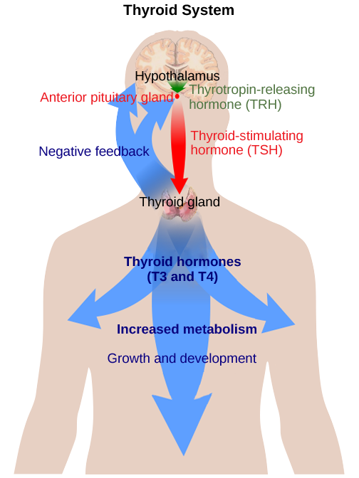
Hormone production and release are primarily controlled by negative feedback, as described in the discussion on homeostasis. In this way, the concentration of hormones in blood is maintained within a narrow range. For example, the anterior pituitary signals the thyroid to release thyroid hormones. Increasing levels of these hormones in the blood then give feedback to the hypothalamus and anterior pituitary to inhibit further signaling to the thyroid gland (Figure 16.14).
Goiter, a disease caused by iodine deficiency, results in the inability of the thyroid gland to form T3 and T4. The body typically attempts to compensate by producing greater amounts of TSH. Which of the following symptoms would you expect goiter to cause?
- Hypothyroidism, resulting in weight gain, cold sensitivity, and reduced mental activity.
- Hyperthyroidism, resulting in weight loss, profuse sweating, and increased heart rate.
- Hyperthyroidism, resulting in weight gain, cold sensitivity, and reduced mental activity.
- Hypothyroidism, resulting in weight loss, profuse sweating, and increased heart rate.
16.5. Musculoskeletal System*
By the end of this section, you will be able to:
- Discuss the axial and appendicular parts of the skeletal system
- Explain the role of joints in skeletal movement
- Explain the role of muscles in locomotion
The muscular and skeletal systems provide support to the body and allow for movement. The bones of the skeleton protect the body’s internal organs and support the weight of the body. The muscles of the muscular system contract and pull on the bones, allowing for movements as diverse as standing, walking, running, and grasping items.
Injury or disease affecting the musculoskeletal system can be very debilitating. The most common musculoskeletal diseases worldwide are caused by malnutrition, which can negatively affect development and maintenance of bones and muscles. Other diseases affect the joints, such as arthritis, which can make movement difficult and, in advanced cases, completely impair mobility.
Progress in the science of prosthesis design has resulted in the development of artificial joints, with joint replacement surgery in the hips and knees being the most common. Replacement joints for shoulders, elbows, and fingers are also available.
Skeletal System
The human skeleton is an endoskeleton that consists of 206 bones in the adult. An endoskeleton develops within the body rather than outside like the exoskeleton of insects. The skeleton has five main functions: providing support to the body, storing minerals and lipids, producing blood cells, protecting internal organs, and allowing for movement. The skeletal system in vertebrates is divided into the axial skeleton (which consists of the skull, vertebral column, and rib cage), and the appendicular skeleton (which consists of limb bones, the pectoral or shoulder girdle, and the pelvic girdle).

Explore the human skeleton by viewing the following video with digital 3D sculpturing.
The axial skeleton forms the central axis of the body and includes the bones of the skull, ossicles of the middle ear, hyoid bone of the throat, vertebral column, and the thoracic cage (rib cage) (Figure 16.15).
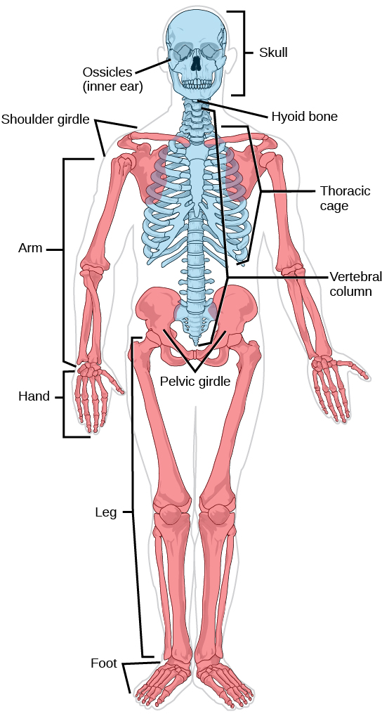
The bones of the skull support the structures of the face and protect the brain. The skull consists of cranial bones and facial bones. The cranial bones form the cranial cavity, which encloses the brain and serves as an attachment site for muscles of the head and neck. In the adult they are tightly jointed with connective tissue and adjoining bones do not move.
The auditory ossicles of the middle ear transmit sounds from the air as vibrations to the fluid-filled cochlea. The auditory ossicles consist of two malleus (hammer) bones, two incus (anvil) bones, and two stapes (stirrups), one on each side. Facial bones provide cavities for the sense organs (eyes, mouth, and nose), and serve as attachment points for facial muscles.
The hyoid bone lies below the mandible in the front of the neck. It acts as a movable base for the tongue and is connected to muscles of the jaw, larynx, and tongue. The mandible forms a joint with the base of the skull. The mandible controls the opening to the mouth and hence, the airway and gut.
The vertebral column, or spinal column, surrounds and protects the spinal cord, supports the head, and acts as an attachment point for ribs and muscles of the back and neck. It consists of 26 bones: the 24 vertebrae, the sacrum, and the coccyx. Each vertebral body has a large hole in the center through which the spinal cord passes down to the level of the first lumbar vertebra. Below this level, the hole contains spinal nerves which exit between the vertebrae. There is a notch on each side of the hole through which the spinal nerves, can exit from the spinal cord to serve different regions of the body. The vertebral column is approximately 70 cm (28 in) in adults and is curved, which can be seen from a side view.
Intervertebral discs composed of fibrous cartilage lie between adjacent vertebrae from the second cervical vertebra to the sacrum. Each disc helps form a slightly moveable joint and acts as a cushion to absorb shocks from movements such as walking and running.
The thoracic cage, also known as the rib cage consists of the ribs, sternum, thoracic vertebrae, and costal cartilages. The thoracic cage encloses and protects the organs of the thoracic cavity including the heart and lungs. It also provides support for the shoulder girdles and upper limbs and serves as the attachment point for the diaphragm, muscles of the back, chest, neck, and shoulders. Changes in the volume of the thorax enable breathing. The sternum, or breastbone, is a long flat bone located at the anterior of the chest. Like the skull, it is formed from many bones in the embryo, which fuse in the adult. The ribs are 12 pairs of long curved bones that attach to the thoracic vertebrae and curve toward the front of the body, forming the ribcage. Costal cartilages connect the anterior ends of most ribs to the sternum.
The appendicular skeleton is composed of the bones of the upper and lower limbs. It also includes the pectoral, or shoulder girdle, which attaches the upper limbs to the body, and the pelvic girdle, which attaches the lower limbs to the body (Figure 16.15).
The pectoral girdle bones transfer force generated by muscles acting on the upper limb to the thorax. It consists of the clavicles (or collarbones) in the anterior, and the scapulae (or shoulder blades) in the posterior.
The upper limb contains bones of the arm (shoulder to elbow), the forearm, and the hand. The humerus is the largest and longest bone of the upper limb. It forms a joint with the shoulder and with the forearm at the elbow. The forearm extends from the elbow to the wrist and consists of two bones. The hand includes the bones of the wrist, the palm, and the bones of the fingers.
The pelvic girdle attaches to the lower limbs of the axial skeleton. Since it is responsible for bearing the weight of the body and for locomotion, the pelvic girdle is securely attached to the axial skeleton by strong ligaments. It also has deep sockets with robust ligaments that securely attach to the femur. The pelvic girdle is mainly composed of two large hip bones. The hip bones join together in the anterior of the body at a joint called the pubic symphysis and with the bones of the sacrum at the posterior of the body.
The lower limb consists of the thigh, the leg, and the foot. The bones of the lower limbs are thicker and stronger than the bones of the upper limbs to support the entire weight of the body and the forces from locomotion. The femur, or thighbone, is the longest, heaviest, and strongest bone in the body. The femur and pelvis form the hip joint. At its other end, the femur, along with the shinbone and kneecap, form the knee joint.
Joints and Skeletal Movement
The point at which two or more bones meet is called a joint, or articulation. Joints are responsible for movement, such as the movement of limbs, and stability, such as the stability found in the bones of the skull.
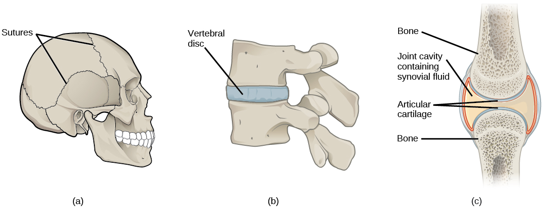
There are two ways to classify joints: based on their structure or based on their function. The structural classification divides joints into fibrous, cartilaginous, and synovial joints depending on the material composing the joint and the presence or absence of a cavity in the joint. The bones of fibrous joints are held together by fibrous connective tissue. There is no cavity, or space, present between the bones, so most fibrous joints do not move at all, or are only capable of minor movements. The joints between the bones in the skull and between the teeth and the bone of their sockets are examples of fibrous joints (Figure 16.16a).
Cartilaginous joints are joints in which the bones are connected by cartilage (Figure 16.16b). An example is found at the joints between vertebrae, the so-called “disks” of the backbone. Cartilaginous joints allow for very little movement.
Synovial joints are the only joints that have a space between the adjoining bones (Figure 16.16c). This space is referred to as the joint cavity and is filled with fluid. The fluid lubricates the joint, reducing friction between the bones and allowing for greater movement. The ends of the bones are covered with cartilage and the entire joint is surrounded by a capsule. Synovial joints are capable of the greatest movement of the joint types. Knees, elbows, and shoulders are examples of synovial joints.
The wide range of movement allowed by synovial joints produces different types of movements. Angular movements are produced when the angle between the bones of a joint changes. Flexion, or bending, occurs when the angle between the bones decreases. Moving the forearm upward at the elbow is an example of flexion. Extension is the opposite of flexion in that the angle between the bones of a joint increases. Rotational movement is the movement of a bone as it rotates around its own longitudinal axis. Movement of the head as in saying “no” is an example of rotation.
Rheumatologists are medical doctors who specialize in the diagnosis and treatment of disorders of the joints, muscles, and bones. They diagnose and treat diseases such as arthritis, musculoskeletal disorders, osteoporosis, plus autoimmune diseases like ankylosing spondylitis, a chronic spinal inflammatory disease and rheumatoid arthritis.
Rheumatoid arthritis (RA) is an inflammatory disorder that primarily affects synovial joints of the hands, feet, and cervical spine. Affected joints become swollen, stiff, and painful. Although it is known that RA is an autoimmune disease in which the body’s immune system mistakenly attacks healthy tissue, the exact cause of RA remains unknown. Immune cells from the blood enter joints and the joint capsule causing cartilage breakdown and swelling of the joint lining. Breakdown of cartilage causes bones to rub against each other causing pain. RA is more common in women than men and the age of onset is usually between 40 to 50 years.
Rheumatologists can diagnose RA based on symptoms such as joint inflammation and pain, x-ray and MRI imaging, and blood tests. Arthrography is a type of medical imaging of joints that uses a contrast agent, such as a dye that is opaque to x-rays. This allows the soft tissue structures of joints—such as cartilage, tendons, and ligaments—to be visualized. An arthrogram differs from a regular x-ray by showing the surface of soft tissues lining the joint in addition to joint bones. An arthrogram allows early degenerative changes in joint cartilage to be detected before bones become affected.
There is currently no cure for RA; however, rheumatologists have a number of treatment options available. Treatments are divided into those that reduce the symptoms of the disease and those that reduce the damage to bone and cartilage caused by the disease. Early stages can be treated with rest of the affected joints through the use of a cane, or with joint splints that minimize inflammation. When inflammation has decreased, exercise can be used to strengthen muscles that surround the joint and to maintain joint flexibility. If joint damage is more extensive, medications can be used to relieve pain and decrease inflammation. Anti-inflammatory drugs that may be used include aspirin, topical pain relievers, and corticosteroid injections. Surgery may be required in cases where joint damage is severe. Physicians are now using drugs that reduce the damage to bones and cartilage caused by the disease to slow its development. These drugs are diverse in their mechanisms but they all act to reduce the impact of the autoimmune response, for example by inhibiting the inflammatory response or reducing the number of T lymphocytes, a cell of the immune system.
Muscles
Muscles allow for movement such as walking, and they also facilitate bodily processes such as respiration and digestion. The body contains three types of muscle tissue: skeletal muscle, cardiac muscle, and smooth muscle (Figure 16.17).
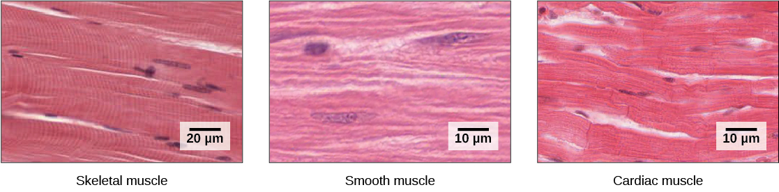
Skeletal muscle tissue forms skeletal muscles, which attach to bones and sometimes the skin and control locomotion and any other movement that can be consciously controlled. Because it can be controlled intentionally, skeletal muscle is also called voluntary muscle. When viewed under a microscope, skeletal muscle tissue has a striped or striated appearance. This appearance results from the arrangement of the proteins inside the cell that are responsible for contraction. The cells of skeletal muscle are long and tapered and have multiple nuclei on the periphery of each cell.
Smooth muscle tissue occurs in the walls of hollow organs such as the intestines, stomach, and urinary bladder, and around passages such as in the respiratory tract and blood vessels. Smooth muscle has no striations, is not under voluntary control, and is called involuntary muscle. Smooth muscle cells have a single nucleus.
Cardiac muscle tissue is only found in the heart. The contractions of cardiac muscle tissue pump blood throughout the body and maintain blood pressure. Like skeletal muscle, cardiac muscle is striated, but unlike skeletal muscle, cardiac muscle cannot be consciously controlled and is called involuntary muscle. The cells of cardiac muscle tissue are connected to each other through intercalated disks and usually have just one nucleus per cell.
Skeletal Muscle Fiber Structure and Function
Each skeletal muscle fiber is a skeletal muscle cell. Within each muscle fiber are myofibrils, long cylindrical structures that lie parallel to the muscle fiber. Myofibrils run the entire length of the muscle fiber. They attach to the plasma membrane, called the sarcolemma, at their ends, so that as myofibrils shorten, the entire muscle cell contracts (Figure 16.18).
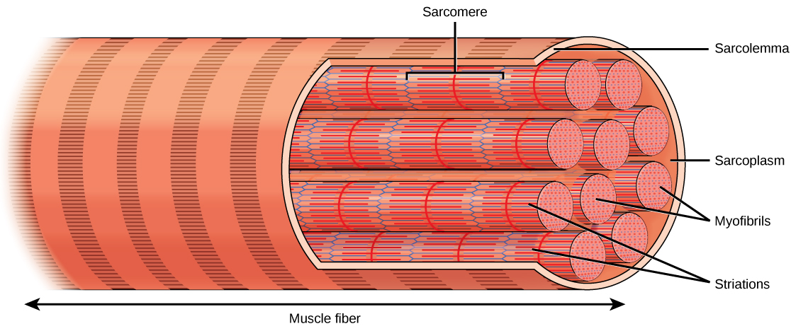
The striated appearance of skeletal muscle tissue is a result of repeating bands of the proteins actin and myosin that occur along the length of myofibrils.
Myofibrils are composed of smaller structures called myofilaments. There are two main types of myofilaments: thick filaments and thin filaments. Thick filaments are composed of the protein myosin. The primary component of thin filaments is the protein actin.
The thick and thin filaments alternate with each other in a structure called a sarcomere. The sarcomere is the unit of contraction in a muscle cell. Contraction is stimulated by an electrochemical signal from a nerve cell associated with the muscle fiber. For a muscle cell to contract, the sarcomere must shorten. However, thick and thin filaments do not shorten. Instead, they slide by one another, causing the sarcomere to shorten while the filaments remain the same length. The sliding is accomplished when a molecular extension of myosin, called the myosin head, temporarily binds to an actin filament next to it and through a change in conformation, bends, dragging the two filaments in opposite directions. The myosin head then releases its actin filament, relaxes, and then repeats the process, dragging the two filaments further along each other. The combined activity of many binding sites and repeated movements within the sarcomere causes it to contract. The coordinated contractions of many sarcomeres in a myofibril leads to contraction of the entire muscle cell and ultimately the muscle itself. The movement of the myosin head requires ATP, which provides the energy for the contraction.

View this animation to see how muscle fibers are organized.
16.6. Nervous System*
By the end of this section, you will be able to:
- Describe the form and function of a neuron
- Describe the basic parts and functions of the central nervous system
- Describe the basic parts and functions of the peripheral nervous system
As you read this, your nervous system is performing several functions simultaneously. The visual system is processing what is seen on the page; the motor system controls your eye movements and the turn of the pages (or click of the mouse); the prefrontal cortex maintains attention. Even fundamental functions, like breathing and regulation of body temperature, are controlled by the nervous system. The nervous system is one of two systems that exert control over all the organ systems of the body; the other is the endocrine system. The nervous system’s control is much more specific and rapid than the hormonal system. It communicates signals through cells and the tiny gaps between them rather than through the circulatory system as in the endocrine system. It uses a combination of chemical and electrochemical signals, rather than purely chemical signals used by the endocrine system to cover long distances quickly. The nervous system acquires information from sensory organs, processes it and then may initiate a response either through motor function, leading to movement, or in a change in the organism’s physiological state.
Nervous systems throughout the animal kingdom vary in structure and complexity. Some organisms, like sea sponges, lack a true nervous system. Others, like jellyfish, lack a true brain and instead have a system of separate but connected nerve cells (neurons) called a “nerve net.” Flatworms have both a central nervous system (CNS), made up of a ganglion (clusters of connected neurons) and two nerve cords, and a peripheral nervous system (PNS) containing a system of nerves that extend throughout the body. The insect nervous system is more complex but also fairly decentralized. It contains a brain, ventral nerve cord, and ganglia. These ganglia can control movements and behaviors without input from the brain.
Compared to invertebrates, vertebrate nervous systems are more complex, centralized, and specialized. While there is great diversity among different vertebrate nervous systems, they all share a basic structure: a CNS that contains a brain and spinal cord and a PNS made up of peripheral sensory and motor nerves. One interesting difference between the nervous systems of invertebrates and vertebrates is that the nerve cords of many invertebrates are located ventrally (toward the stomach) whereas the vertebrate spinal cords are located dorsally (toward the back). There is debate among evolutionary biologists as to whether these different nervous system plans evolved separately or whether the invertebrate body plan arrangement somehow “flipped” during the evolution of vertebrates.
The nervous system is made up of neurons, specialized cells that can receive and transmit chemical or electrical signals, and glia, cells that provide support functions for the neurons. There is great diversity in the types of neurons and glia that are present in different parts of the nervous system.
Neurons and Glial Cells
The nervous system of the common laboratory fly, Drosophila melanogaster, contains around 100,000 neurons, the same number as a lobster. This number compares to 75 million in the mouse and 300 million in the octopus. A human brain contains around 86 billion neurons. Despite these very different numbers, the nervous systems of these animals control many of the same behaviors—from basic reflexes to more complicated behaviors like finding food and courting mates. The ability of neurons to communicate with each other as well as with other types of cells underlies all of these behaviors.
Most neurons share the same cellular components. But neurons are also highly specialized—different types of neurons have different sizes and shapes that relate to their functional roles.
Like other cells, each neuron has a cell body (or soma) that contains a nucleus, smooth and rough endoplasmic reticulum, Golgi apparatus, mitochondria, and other cellular components. Neurons also contain unique structures for receiving and sending the electrical signals that make communication between neurons possible (Figure 16.19). Dendrites are tree-like structures that extend away from the cell body to receive messages from other neurons at specialized junctions called synapses. Although some neurons do not have any dendrites, most have one or many dendrites.
The bilayer lipid membrane that surrounds a neuron is impermeable to ions. To enter or exit the neuron, ions must pass through ion channels that span the membrane. Some ion channels need to be activated to open and allow ions to pass into or out of the cell. These ion channels are sensitive to the environment and can change their shape accordingly. Ion channels that change their structure in response to voltage changes are called voltage-gated ion channels. The difference in total charge between the inside and outside of the cell is called the membrane potential.
A neuron at rest is negatively charged: the inside of a cell is approximately 70 millivolts more negative than the outside (–70 mV). This voltage is called the resting membrane potential; it is caused by differences in the concentrations of ions inside and outside the cell and the selective permeability created by ion channels. Sodium-potassium pumps in the membrane produce the different ion concentrations inside and outside of the cell by bringing in two K+ ions and removing three Na+ ions. The actions of this pump are costly: one molecule of ATP is used up for each turn. Up to 50 percent of a neuron’s ATP is used in maintaining its membrane resting potential. Potassium ions (K+), which are higher inside the cell, move fairly freely out of the neuron through potassium channels; this loss of positive charge produces a net negative charge inside the cell. Sodium ions (Na+), which are low inside, have a driving force to enter but move less freely. Their channels are voltage dependent and will open when a slight change in the membrane potential triggers them.
A neuron can receive input from other neurons and, if this input is strong enough, send the signal to downstream neurons. Transmission of a signal between neurons is generally carried by a chemical, called a neurotransmitter, which diffuses from the axon of one neuron to the dendrite of a second neuron. When neurotransmitter molecules bind to receptors located on a neuron’s dendrites, the neurotransmitter opens ion channels in the dendrite’s plasma membrane. This opening allows sodium ions to enter the neuron and results in depolarization of the membrane—a decrease in the voltage across the neuron membrane. Once a signal is received by the dendrite, it then travels passively to the cell body. A large enough signal from neurotransmitters will reach the axon. If it is strong enough (that is, if the threshold of excitation, a depolarization to around –60mV is reached), then depolarization creates a positive feedback loop: as more Na+ ions enter the cell, the axon becomes further depolarized, opening even more sodium channels at further distances from the cell body. This will cause voltage dependent Na+ channels further down the axon to open and more positive ions to enter the cell. In the axon, this “signal” will become a self-propagating brief reversal of the resting membrane potential called an action potential.
An action potential is an all-or-nothing event; it either happens or it does not. The threshold of excitation must be reached for the neuron to “fire” an action potential. As sodium ions rush into the cell, depolarization actually reverses the charge across the membrane form -70mv to +30mV. This change in the membrane potential causes voltage-gated K+ channels to open, and K+ begins to leave the cell, repolarizing it. At the same time, Na+ channels inactivate so no more Na+ enters the cell. K+ ions continue to leave the cell and the membrane potential returns to the resting potential. At the resting potential, the K+ channels close and Na+ channels reset. The depolarization of the membrane proceeds in a wave down the length of the axon. It travels in only one direction because the sodium channels have been inactivated and unavailable until the membrane potential is near the resting potential again; at this point they are reset to closed and can be opened again.
An axon is a tube-like structure that propagates the signal from the cell body to specialized endings called axon terminals. These terminals in turn then synapse with other neurons, muscle, or target organs. When the action potential reaches the axon terminal, this causes the release of neurotransmitter onto the dendrite of another neuron. Neurotransmitters released at axon terminals allow signals to be communicated to these other cells, and the process begins again. Neurons usually have one or two axons, but some neurons do not contain any axons.
Some axons are covered with a special structure called a myelin sheath, which acts as an insulator to keep the electrical signal from dissipating as it travels down the axon. This insulation is important, as the axon from a human motor neuron can be as long as a meter (3.2 ft)—from the base of the spine to the toes. The myelin sheath is produced by glial cells. Along the axon there are periodic gaps in the myelin sheath. These gaps are called nodes of Ranvier and are sites where the signal is “recharged” as it travels along the axon.
It is important to note that a single neuron does not act alone—neuronal communication depends on the connections that neurons make with one another (as well as with other cells, like muscle cells). Dendrites from a single neuron may receive synaptic contact from many other neurons. For example, dendrites from a Purkinje cell in the cerebellum are thought to receive contact from as many as 200,000 other neurons.
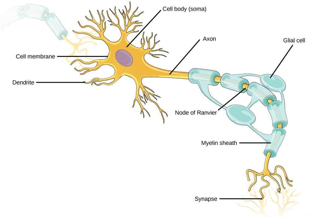
At one time, scientists believed that people were born with all the neurons they would ever have. Research performed during the last few decades indicates that neurogenesis, the birth of new neurons, continues into adulthood. Neurogenesis was first discovered in songbirds that produce new neurons while learning songs. For mammals, new neurons also play an important role in learning: about 1,000 new neurons develop in the hippocampus (a brain structure involved in learning and memory) each day. While most of the new neurons will die, researchers found that an increase in the number of surviving new neurons in the hippocampus correlated with how well rats learned a new task. Interestingly, both exercise and some antidepressant medications also promote neurogenesis in the hippocampus. Stress has the opposite effect. While neurogenesis is quite limited compared to regeneration in other tissues, research in this area may lead to new treatments for disorders such as Alzheimer’s, stroke, and epilepsy.
How do scientists identify new neurons? A researcher can inject a compound called bromodeoxyuridine (BrdU) into the brain of an animal. While all cells will be exposed to BrdU, BrdU will only be incorporated into the DNA of newly generated cells that are in S phase. A technique called immunohistochemistry can be used to attach a fluorescent label to the incorporated BrdU, and a researcher can use fluorescent microscopy to visualize the presence of BrdU, and thus new neurons, in brain tissue (Figure 16.20).
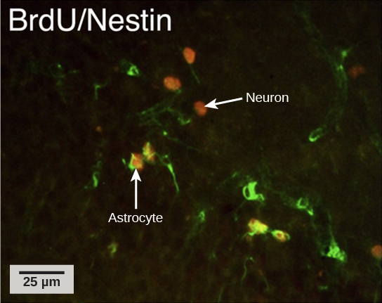

Visit this link interactive lab to see more information about neurogenesis, including an interactive laboratory simulation and a video that explains how BrdU labels new cells.
While glial cells are often thought of as the supporting cast of the nervous system, the number of glial cells in the brain actually outnumbers the number of neurons by a factor of 10. Neurons would be unable to function without the vital roles that are fulfilled by these glial cells. Glia guide developing neurons to their destinations, buffer ions and chemicals that would otherwise harm neurons, and provide myelin sheaths around axons. When glia do not function properly, the result can be disastrous—most brain tumors are caused by mutations in glia.
How Neurons Communicate
All functions performed by the nervous system—from a simple motor reflex to more advanced functions like making a memory or a decision—require neurons to communicate with one another. Neurons communicate between the axon of one neuron and the dendrites, and sometimes the cell body, of another neuron across the gap between them, known as the synaptic cleft. When an action potential reaches the end of an axon it stimulates the release of neurotransmitter molecules into the synaptic cleft between the synaptic knob of the axon and the post-synaptic membrane of the dendrite or soma of the next cell. The neurotransmitter is released through exocytosis of vesicles containing the neurotransmitter molecules. The neurotransmitter diffuses across the synaptic cleft and binds to receptors in the post-synaptic membrane. These receptor molecules are chemically regulated ion channels and will open, allowing sodium to enter the cell. If sufficient neurotransmitter has been released an action potential may be initiated in the next cell, but this is not guaranteed. If insufficient neurotransmitter is released the nerve signal will die at this point. There are a number of different neurotransmitters that are specific to neuron types that have specific functions.
The Central Nervous System
The central nervous system (CNS) is made up of the brain and spinal cord and is covered with three layers of protective coverings called meninges (“meninges” is derived from the Greek and means “membranes”) (Figure 16.21). The outermost layer is the dura mater, the middle layer is the web-like arachnoid mater, and the inner layer is the pia mater, which directly contacts and covers the brain and spinal cord. The space between the arachnoid and pia maters is filled with cerebrospinal fluid (CSF). The brain floats in CSF, which acts as a cushion and shock absorber.
The Brain
The brain is the part of the central nervous system that is contained in the cranial cavity of the skull. It includes the cerebral cortex, limbic system, basal ganglia, thalamus, hypothalamus, cerebellum, brainstem, and retinas. The outermost part of the brain is a thick piece of nervous system tissue called the cerebral cortex. The cerebral cortex, limbic system, and basal ganglia make up the two cerebral hemispheres. A thick fiber bundle called the corpus callosum (corpus = “body”; callosum = “tough”) connects the two hemispheres. Although there are some brain functions that are localized more to one hemisphere than the other, the functions of the two hemispheres are largely redundant. In fact, sometimes (very rarely) an entire hemisphere is removed to treat severe epilepsy. While patients do suffer some deficits following the surgery, they can have surprisingly few problems, especially when the surgery is performed on children who have very immature nervous systems.
In other surgeries to treat severe epilepsy, the corpus callosum is cut instead of removing an entire hemisphere. This causes a condition called split-brain, which gives insights into unique functions of the two hemispheres. For example, when an object is presented to patients’ left visual field, they may be unable to verbally name the object (and may claim to not have seen an object at all). This is because the visual input from the left visual field crosses and enters the right hemisphere and cannot then signal to the speech center, which generally is found in the left side of the brain. Remarkably, if a split-brain patient is asked to pick up a specific object out of a group of objects with the left hand, the patient will be able to do so but will still be unable to verbally identify it.

Visit the following website to learn more about split-brain patients and to play a game where you can model split-brain experiments yourself.
Each hemisphere contains regions called lobes that are involved in different functions. Each hemisphere of the mammalian cerebral cortex can be broken down into four functionally and spatially defined lobes: frontal, parietal, temporal, and occipital (Figure 16.22).
The frontal lobe is located at the front of the brain, over the eyes. This lobe contains the olfactory bulb, which processes smells. The frontal lobe also contains the motor cortex, which is important for planning and implementing movement. Areas within the motor cortex map to different muscle groups. Neurons in the frontal lobe also control cognitive functions like maintaining attention, speech, and decision-making. Studies of humans who have damaged their frontal lobes show that parts of this area are involved in personality, socialization, and assessing risk. The parietal lobe is located at the top of the brain. Neurons in the parietal lobe are involved in speech and also reading. Two of the parietal lobe’s main functions are processing somatosensation—touch sensations like pressure, pain, heat, cold—and processing proprioception—the sense of how parts of the body are oriented in space. The parietal lobe contains a somatosensory map of the body similar to the motor cortex. The occipital lobe is located at the back of the brain. It is primarily involved in vision—seeing, recognizing, and identifying the visual world. The temporal lobe is located at the base of the brain and is primarily involved in processing and interpreting sounds. It also contains the hippocampus (named from the Greek for “seahorse,” which it resembles in shape) a structure that processes memory formation. The role of the hippocampus in memory was partially determined by studying one famous epileptic patient, HM, who had both sides of his hippocampus removed in an attempt to cure his epilepsy. His seizures went away, but he could no longer form new memories (although he could remember some facts from before his surgery and could learn new motor tasks).
Interconnected brain areas called the basal ganglia play important roles in movement control and posture. The basal ganglia also regulate motivation.
The thalamus acts as a gateway to and from the cortex. It receives sensory and motor inputs from the body and also receives feedback from the cortex. This feedback mechanism can modulate conscious awareness of sensory and motor inputs depending on the attention and arousal state of the animal. The thalamus helps regulate consciousness, arousal, and sleep states.
Below the thalamus is the hypothalamus. The hypothalamus controls the endocrine system by sending signals to the pituitary gland. Among other functions, the hypothalamus is the body’s thermostat—it makes sure the body temperature is kept at appropriate levels. Neurons within the hypothalamus also regulate circadian rhythms, sometimes called sleep cycles.
The limbic system is a connected set of structures that regulates emotion, as well as behaviors related to fear and motivation. It plays a role in memory formation and includes parts of the thalamus and hypothalamus as well as the hippocampus. One important structure within the limbic system is a temporal lobe structure called the amygdala. The two amygdala (one on each side) are important both for the sensation of fear and for recognizing fearful faces.
The cerebellum (cerebellum = “little brain”) sits at the base of the brain on top of the brainstem. The cerebellum controls balance and aids in coordinating movement and learning new motor tasks. The cerebellum of birds is large compared to other vertebrates because of the coordination required by flight.
The brainstem connects the rest of the brain with the spinal cord and regulates some of the most important and basic functions of the nervous system including breathing, swallowing, digestion, sleeping, walking, and sensory and motor information integration.
Spinal cord
Connecting to the brainstem and extending down the body through the spinal column is the spinal cord. The spinal cord is a thick bundle of nerve tissue that carries information about the body to the brain and from the brain to the body. The spinal cord is contained within the meninges and the bones of the vertebral column but is able to communicate signals to and from the body through its connections with spinal nerves (part of the peripheral nervous system). A cross-section of the spinal cord looks like a white oval containing a gray butterfly-shape (Figure 16.23). Axons make up the “white matter” and neuron and glia cell bodies (and interneurons) make up the “gray matter.” Axons and cell bodies in the dorsa spinal cord convey mostly sensory information from the body to the brain. Axons and cell bodies in the spinal cord primarily transmit signals controlling movement from the brain to the body.
The spinal cord also controls motor reflexes. These reflexes are quick, unconscious movements—like automatically removing a hand from a hot object. Reflexes are so fast because they involve local synaptic connections. For example, the knee reflex that a doctor tests during a routine physical is controlled by a single synapse between a sensory neuron and a motor neuron. While a reflex may only require the involvement of one or two synapses, synapses with interneurons in the spinal column transmit information to the brain to convey what happened (the knee jerked, or the hand was hot).
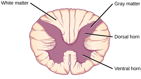
The Peripheral Nervous System
The peripheral nervous system (PNS) is the connection between the central nervous system and the rest of the body. The PNS can be broken down into the autonomic nervous system, which controls bodily functions without conscious control, and the sensory-somatic nervous system, which transmits sensory information from the skin, muscles, and sensory organs to the CNS and sends motor commands from the CNS to the muscles.
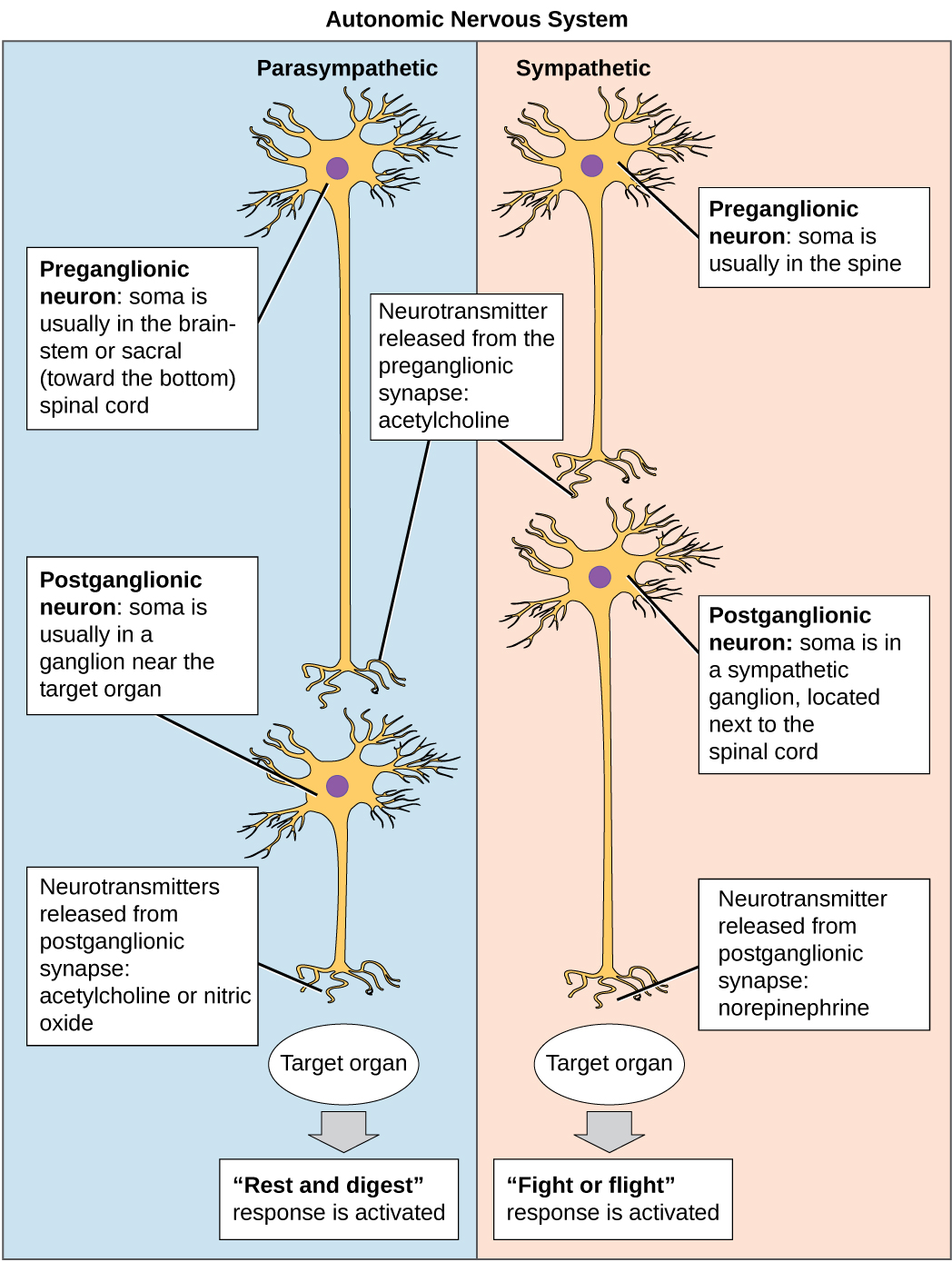
The autonomic nervous system serves as the relay between the CNS and the internal organs. It controls the lungs, the heart, smooth muscle, and exocrine and endocrine glands. The autonomic nervous system controls these organs largely without conscious control; it can continuously monitor the conditions of these different systems and implement changes as needed. Signaling to the target tissue usually involves two synapses: a preganglionic neuron (originating in the CNS) synapses to a neuron in a ganglion that, in turn, synapses on the target organ (Figure 16.24). There are two divisions of the autonomic nervous system that often have opposing effects: the sympathetic nervous system and the parasympathetic nervous system.
The sympathetic nervous system is responsible for the immediate responses an animal makes when it encounters a dangerous situation. One way to remember this is to think of the “fight-or-flight” response a person feels when encountering a snake (“snake” and “sympathetic” both begin with “s”). Examples of functions controlled by the sympathetic nervous system include an accelerated heart rate and inhibited digestion. These functions help prepare an organism’s body for the physical strain required to escape a potentially dangerous situation or to fend off a predator.
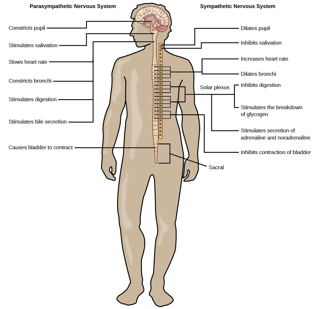
While the sympathetic nervous system is activated in stressful situations, the parasympathetic nervous system allows an animal to “rest and digest.” One way to remember this is to think that during a restful situation like a picnic, the parasympathetic nervous system is in control (“picnic” and “parasympathetic” both start with “p”). Parasympathetic preganglionic neurons have cell bodies located in the brainstem and in the sacral (toward the bottom) spinal cord (Figure 16.25). The parasympathetic nervous system resets organ function after the sympathetic nervous system is activated including slowing of heart rate, lowered blood pressure, and stimulation of digestion.
The sensory-somatic nervous system is made up of cranial and spinal nerves and contains both sensory and motor neurons. Sensory neurons transmit sensory information from the skin, skeletal muscle, and sensory organs to the CNS. Motor neurons transmit messages about desired movement from the CNS to the muscles to make them contract. Without its sensory-somatic nervous system, an animal would be unable to process any information about its environment (what it sees, feels, hears, and so on) and could not control motor movements. Unlike the autonomic nervous system, which usually has two synapses between the CNS and the target organ, sensory and motor neurons usually have only one synapse—one ending of the neuron is at the organ and the other directly contacts a CNS neuron.
Glossary
- action potential
- a momentary change in the electrical potential of a neuron (or muscle) membrane
- adrenal gland
- the endocrine gland associated with the kidneys
- alveolus
- (plural: alveoli) (also, air sacs) the terminal structure of the lung passage where gas exchange occurs
- amygdala
- a structure within the limbic system that processes fear
- amylase
- an enzyme found in saliva and secreted by the pancreas that converts carbohydrates to maltose
- anus
- the exit point of the digestive system for waste material
- aorta
- the major artery that takes blood away from the heart to the systemic circulatory system
- appendicular skeleton
- the skeleton composed of the bones of the upper limbs, which function to grasp and manipulate objects, and the lower limbs, which permit locomotion
- artery
- a blood vessel that takes blood away from the heart
- atrium
- (plural: atria) a chamber of the heart that receives blood from the veins
- auditory ossicles
- (also, middle ear bones) the bones that transduce sounds from the air into vibrations in the fluid-filled cochlea
- autonomic nervous system
- the part of the peripheral nervous system that controls bodily functions
- axial skeleton
- skeleton that forms the central axis of the body and includes the bones of the skull, the ossicles of the middle ear, the hyoid bone of the throat, the vertebral column, and the thoracic cage (ribcage)
- axon
- a tube-like structure that propagates a signal from a neuron’s cell body to axon terminals
- basal ganglia
- an interconnected collections of cells in the brain that are involved in movement and motivation
- bicuspid valve
- a one-way opening between the atrium and the ventricle in the left side of the heart
- bile
- a digestive juice produced by the liver; important for digestion of lipids
- bolus
- a mass of food resulting from chewing action and wetting by saliva
- brainstem
- a portion of brain that connects with the spinal cord; controls basic nervous system functions like breathing and swallowing
- bronchi
- (singular: bronchus) smaller branches of cartilaginous tissue that stem off of the trachea; air is funneled through the bronchi to the region where gas exchange occurs in the alveoli
- bronchiole
- an airway that extends from the main bronchus to the alveolar sac
- capillary
- the smallest blood vessel that allows the passage of individual blood cells and the site of diffusion of oxygen and nutrient exchange
- cardiac cycle
- the filling and emptying the heart of blood caused by electrical signals that cause the heart muscles to contract and relax
- cardiac muscle tissue
- the muscle tissue found only in the heart; cardiac contractions pump blood throughout the body and maintain blood pressure
- cartilaginous joint
- a joint in which the bones are connected by cartilage
- central nervous system (CNS)
- the nervous system made up of the brain and spinal cord; covered with three layers of protective meninges
- cerebellum
- the brain structure involved in posture, motor coordination, and learning new motor actions
- cerebral cortex
- the outermost sheet of brain tissue; involved in many higher-order functions
- cerebrospinal fluid (CSF)
- a clear liquid that surrounds the brain and fills its ventricles and acts as a shock absorber
- chyme
- a mixture of partially digested food and stomach juices
- closed circulatory system
- a system that has the blood separated from the bodily interstitial fluid and contained in blood vessels
- colon
- the largest portion of the large intestine consisting of the ascending colon, transverse colon, and descending colon
- corpus callosum
- a thick nerve bundle that connects the cerebral hemispheres
- dendrite
- a structure that extends away from the cell body to receive messages from other neurons
- depolarization
- a change in the membrane potential to a less negative value
- diaphragm
- a skeletal muscle located under lungs that encloses the lungs in the thorax
- diastole
- the relaxation phase of the cardiac cycle when the heart is relaxed and the ventricles are filling with blood
- down-regulation
- a decrease in the number of hormone receptors in response to increased hormone levels
- ectotherm
- an organism that relies primarily on environmental heat sources to maintain its body temperature
- electrocardiogram (ECG)
- a recording of the electrical impulses of the cardiac muscle
- endocrine gland
- the gland that secretes hormones into the surrounding interstitial fluid, which then diffuse into blood and are carried to various organs and tissues within the body
- endotherm
- an organism that relies primarily on internal heat sources to maintain its body temperature
- esophagus
- a tubular organ that connects the mouth to the stomach
- essential nutrient
- a nutrient that cannot be synthesized by the body; it must be obtained from food
- exocrine gland
- the gland that secretes chemicals through ducts that lead to skin surfaces, body cavities, and organ cavities.
- fibrous joint
- a joint held together by fibrous connective tissue
- frontal lobe
- the part of the cerebral cortex that contains the motor cortex and areas involved in planning, attention, and language
- gallbladder
- the organ that stores and concentrates bile
- glia
- (also, glial cells) the cells that provide support functions for neurons
- hippocampus
- the brain structure in the temporal lobe involved in processing memories
- hormone
- a chemical released by cells in one area of the body that affects cells in other parts of the body
- hyoid bone
- the bone that lies below the mandible in the front of the neck
- hypothalamus
- the brain structure that controls hormone release and body homeostasis
- inferior vena cava
- the major vein of the body returning blood from the lower parts of the body to the right atrium
- interstitial fluid
- the fluid found between cells in the body, similar in constitution to the fluid component of blood, but without the high concentrations of proteins
- intracellular hormone receptor
- a hormone receptor in the cytoplasm or nucleus of a cell
- joint
- the point at which two or more bones meet
- kidney
- the organ that performs excretory and osmoregulatory functions
- large intestine
- a digestive system organ that reabsorbs water from undigested material and processes waste matter
- larynx
- the voice box, located within the throat
- limbic system
- a connected brain area that processes emotion and motivation
- liver
- an organ that produces bile for digestion and processes vitamins and lipids
- membrane potential
- a difference in electrical potential between the inside and outside of a cell
- meninges
- (singular: meninx) the membranes that cover and protect the central nervous system
- mineral
- an inorganic, elemental molecule that carries out important roles in the body
- myelin sheath
- a cellular extension containing a fatty substance produced by glia that surrounds and insulates axons
- myofibril
- the long cylindrical structures that lie parallel to the muscle fiber
- myofilament
- the small structures that make up myofibrils
- nasal cavity
- an opening of the respiratory system to the outside environment
- nephron
- the functional unit of the kidney
- neuron
- a specialized cell that can receive and transmit electrical and chemical signals
- occipital lobe
- the part of the cerebral cortex that contains visual cortex and processes visual stimuli
- open circulatory system
- a circulatory system that has the blood mixed with interstitial fluid in the body cavity and directly bathes the organs
- oral cavity
- the point of entry of food into the digestive system
- osmoregulation
- the mechanism by which water and solute concentrations are maintained at desired levels
- osmotic balance
- the appropriate values of water and solute concentrations for a healthy organism
- pancreas
- a gland that secretes digestive juices
- pancreas
- the organ located between the stomach and the small intestine that contains exocrine and endocrine cells
- parasympathetic nervous system
- the division of autonomic nervous system that regulates visceral functions during relaxation
- parathyroid gland
- the gland located on the surface of the thyroid that produces parathyroid hormone
- parietal lobe
- the part of the cerebral cortex involved in processing touch and the sense of the body in space
- pectoral girdle
- the bones that transmit the force generated by the upper limbs to the axial skeleton
- pelvic girdle
- the bones that transmit the force generated by the lower limbs to the axial skeleton
- pepsin
- an enzyme found in the stomach whose main role is protein digestion
- peripheral nervous system (PNS)
- the nervous system that serves as the connection between the central nervous system and the rest of the body; consists of the autonomic nervous system and the sensory-somatic nervous system
- peristalsis
- wave-like movements of muscle tissue
- pharynx
- the throat
- pituitary gland
- the endocrine gland located at the base of the brain composed of an anterior and posterior region; also called hypophysis
- primary bronchus
- (also, main bronchus) a region of the airway within the lung that attaches to the trachea and bifurcates to form the bronchioles
- pulmonary circulation
- the flow of blood away from the heart through the lungs where oxygenation occurs and then back to the heart
- rectum
- the area of the body where feces is stored until elimination
- renal artery
- the artery that delivers blood to the kidney
- renal vein
- the vein that drains blood from the kidney
- salivary gland
- one of three pairs of exocrine glands in the mammalian mouth that secretes saliva, a mix of watery mucus and enzymes
- sarcolemma
- the plasma membrane of a skeletal muscle fiber
- sarcomere
- the functional unit of skeletal muscle
- sensory-somatic nervous system
- the system of sensory and motor nerves
- set point
- the target value of a physiological state in homeostasis
- skeletal muscle tissue
- forms skeletal muscles, which attach to bones and control locomotion and any movement that can be consciously controlled
- skull
- the bone that supports the structures of the face and protects the brain
- small intestine
- the organ where digestion of protein, fats, and carbohydrates is completed
- smooth muscle tissue
- the muscle that occurs in the walls of hollow organs such as the intestines, stomach, and urinary bladder, and around passages such as the respiratory tract and blood vessels
- spinal cord
- a thick fiber bundle that connects the brain with peripheral nerves; transmits sensory and motor information; contains neurons that control motor reflexes
- stomach
- a saclike organ containing acidic digestive juices
- superior vena cava
- the major vein of the body returning blood from the upper part of the body to the right atrium
- sympathetic nervous system
- the division of autonomic nervous system activated during stressful “fight-or-flight” situations
- synapse
- a junction between two neurons where neuronal signals are communicated
- synaptic cleft
- a space between the presynaptic and postsynaptic membranes
- synovial joints
- the only joints that have a space between the adjoining bones
- systemic circulation
- the flow of blood away from the heart to the brain, liver, kidneys, stomach, and other organs, the limbs, and the muscles of the body, and then back to the heart
- systole
- the contraction phase of cardiac cycle when the ventricles are pumping blood into the arteries
- temporal lobe
- the part of the cerebral cortex that processes auditory input; parts of the temporal lobe are involved in speech, memory, and emotion processing
- thalamus
- the brain area that relays sensory information to the cortex
- thoracic cage
- (also, ribcage) the skeleton of the chest, which consists of the ribs, thoracic vertebrae, sternum, and costal cartilages
- threshold of excitation
- the level of depolarization needed for an action potential to fire
- thymus
- the gland located behind the sternum that produces thymosin hormones that contribute to the development of the immune system
- thyroid gland
- an endocrine gland located in the neck that produces thyroid hormones thyroxine and triiodothyronine
- trachea
- the cartilaginous tube that transports air from the throat to the lungs
- tricuspid valve
- a one-way opening between the atrium and the ventricle in the right side of the heart
- up-regulation
- an increase in the number of hormone receptors in response to increased hormone levels
- ureter
- the urine-bearing tubes coming out of the kidney
- urethra
- the tube that conducts urine from the urinary bladder to the external environment
- urinary bladder
- the structure that the ureters empty the urine into
- vein
- a blood vessel that brings blood back to the heart
- ventricle
- (of the heart) a large chamber of the heart that pumps blood into arteries
- vertebral column
- (also, spine) the column that surrounds and protects the spinal cord, supports the head, and acts as an attachment point for ribs and muscles of the back and neck
- vitamin
- an organic substance necessary in small amounts to sustain life
<!–CNX: Start Area: “Sections Summary”–>
Homeostasis is a dynamic equilibrium that is maintained in body tissues and organs. It is dynamic because it is constantly adjusting to the changes that the systems encounter. It is an equilibrium because body functions are kept within a normal range, with some fluctuations around a set point. The kidneys are the main osmoregulatory organs in mammalian systems; they function to filter blood and maintain the dissolved ion concentrations of body fluids. They are made up internally of three distinct regions—the cortex, medulla, and pelvis. The blood vessels that transport blood into and out of the kidneys arise from and merge with the aorta and inferior vena cava, respectively. The nephron is the functional unit of the kidney, which actively filters blood and generates urine. The urine leaves the kidney through the ureter and is stored in the urinary bladder. Urine is voided from the body through the urethra.
There are many organs that work together to digest food and absorb nutrients. The mouth is the point of ingestion and the location where both mechanical and chemical breakdown of food begins. Saliva contains an enzyme called amylase that breaks down carbohydrates. The food bolus travels through the esophagus by peristaltic movements to the stomach. The stomach has an extremely acidic environment. The enzyme pepsin digests protein in the stomach. Further digestion and absorption take place in the small intestine. The large intestine reabsorbs water from the undigested food and stores waste until elimination.
Carbohydrates, proteins, and fats are the primary components of food. Some essential nutrients are required for cellular function but cannot be produced by the animal body. These include vitamins, minerals, some fatty acids, and some amino acids. Food intake in more than necessary amounts is stored as glycogen in the liver and muscle cells, and in adipose tissue. Excess adipose storage can lead to obesity and serious health problems.
Animal respiratory systems are designed to facilitate gas exchange. In mammals, air is warmed and humidified in the nasal cavity. Air then travels down the pharynx and larynx, through the trachea, and into the lungs. In the lungs, air passes through the branching bronchi, reaching the respiratory bronchioles. The respiratory bronchioles open up into the alveolar ducts, alveolar sacs, and alveoli. Because there are so many alveoli and alveolar sacs in the lung, the surface area for gas exchange is very large.
The mammalian circulatory system is a closed system with double circulation passing through the lungs and the body. It consists of a network of vessels containing blood that circulates because of pressure differences generated by the heart.
The heart contains two pumps that move blood through the pulmonary and systemic circulations. There is one atrium and one ventricle on the right side and one atrium and one ventricle on the left side. The pumping of the heart is a function of cardiomyocytes, distinctive muscle cells that are striated like skeletal muscle but pump rhythmically and involuntarily like smooth muscle. The signal for contraction begins in the wall of the right atrium. The electrochemical signal causes the two atria to contract in unison; then the signal causes the ventricles to contract. The blood from the heart is carried through the body by a complex network of blood vessels; arteries take blood away from the heart, and veins bring blood back to the heart.
Hormones cause cellular changes by binding to receptors on or in target cells. The number of receptors on a target cell can increase or decrease in response to hormone activity.
Hormone levels are primarily controlled through negative feedback, in which rising levels of a hormone inhibit its further release.
The pituitary gland is located at the base of the brain. The anterior pituitary receives signals from the hypothalamus and produces six hormones. The posterior pituitary is an extension of the brain and releases hormones (antidiuretic hormone and oxytocin) produced by the hypothalamus. The thyroid gland is located in the neck and is composed of two lobes. The thyroid produces the hormones thyroxine and triiodothyronine. The thyroid also produces calcitonin. The parathyroid glands lie on the posterior surface of the thyroid gland and produce parathyroid hormone.
The adrenal glands are located on top of the kidneys and consist of the adrenal cortex and adrenal medulla. The adrenal cortex produces the corticosteroids, glucocorticoids and mineralocorticoids. The adrenal medulla is the inner part of the adrenal gland and produces epinephrine and norepinephrine.
The pancreas lies in the abdomen between the stomach and the small intestine. Clusters of endocrine cells in the pancreas form the islets of Langerhans, which contain alpha cells that release glucagon and beta cells that release insulin. Some organs possess endocrine activity as a secondary function but have another primary function. The heart produces the hormone atrial natriuretic peptide, which functions to reduce blood volume, pressure, and Na+ concentration. The gastrointestinal tract produces various hormones that aid in digestion. The kidneys produce erythropoietin. The thymus produces hormones that aid in the development of the immune system. The gonads produce steroid hormones, including testosterone in males and estrogen and progesterone in females. Adipose tissue produces leptin, which promotes satiety signals in the brain.
The human skeleton is an endoskeleton that is composed of the axial and appendicular skeleton. The axial skeleton is composed of the bones of the skull, ossicles of the ear, hyoid bone, vertebral column, and ribcage. The skull consists of eight cranial bones and 14 facial bones. Six bones make up the ossicles of the middle ear, while the hyoid bone is located in the neck under the mandible. The vertebral column contains 26 bones and surrounds and protects the spinal cord. The thoracic cage consists of the sternum, ribs, thoracic vertebrae, and costal cartilages. The appendicular skeleton is made up of the upper and lower limbs. The pectoral girdle is composed of the clavicles and the scapulae. The upper limb contains 30 bones in the arm, the forearm, and the hand. The pelvic girdle attaches the lower limbs to the axial skeleton. The lower limb includes the bones of the thigh, the leg, and the foot.
The structural classification of joints divides them into fibrous, cartilaginous, and synovial joints. The bones of fibrous joints are held together by fibrous connective tissue. Cartilaginous joints are joints in which the bones are connected by cartilage. Synovial joints are joints that have a space between the adjoining bones. The movement of synovial joints includes angular and rotational. Angular movements are produced when the angle between the bones of a joint changes. Rotational movement is the movement of a bone as it rotates around its own longitudinal axis.
The body contains three types of muscle tissue: skeletal muscle, cardiac muscle, and smooth muscle. Muscles are composed of individual cells called muscle fibers. Muscle fibers consist of myofilaments composed of the proteins actin and myosin arranged in units called sarcomeres. Contraction of the muscle occurs by the combined action of myosin and actin fibers sliding past each other when the myosin heads bind to the actin fiber, bend, disengage, and then repeat the process.
The nervous system is made up of neurons and glia. Neurons are specialized cells that are capable of sending electrical as well as chemical signals. Most neurons contain dendrites, which receive these signals, and axons that send signals to other neurons or tissues. Glia are non-neuronal cells in the nervous system that support neuronal development and signaling. There are several types of glia that serve different functions.
Neurons have a resting potential across their membranes and when they are stimulated by a strong enough signal from another neuron an action potential may carry an electrochemical signal along the neuron to a synapse with another neuron. Neurotransmitters carry signals across synapses to initiate a response in another neuron.
The vertebrate central nervous system contains the brain and the spinal cord, which are covered and protected by three meninges. The brain contains structurally and functionally defined regions. In mammals, these include the cortex (which can be broken down into four primary functional lobes: frontal, temporal, occipital, and parietal), basal ganglia, thalamus, hypothalamus, limbic system, cerebellum, and brainstem—although structures in some of these designations overlap. While functions may be primarily localized to one structure in the brain, most complex functions, like language and sleep, involve neurons in multiple brain regions. The spinal cord is the information superhighway that connects the brain with the rest of the body through its connections with peripheral nerves. It transmits sensory and motor input and also controls motor reflexes.
The peripheral nervous system contains both the autonomic and sensory-somatic nervous systems. The autonomic nervous system provides unconscious control over visceral functions and has two divisions: the sympathetic and parasympathetic nervous systems. The sympathetic nervous system is activated in stressful situations to prepare the animal for a “fight-or-flight” response. The parasympathetic nervous system is active during restful periods. The sensory-somatic nervous system is made of cranial and spinal nerves that transmit sensory information from skin and muscle to the CNS and motor commands from the CNS to the muscles.
<!–CNX: Start Area: “Art Connections”–>
Figure 16.2 Pyrogens increase body temperature by causing the blood vessels to constrict, inducing shivering, and stopping sweat glands from secreting fluid.
Figure 16.7 Which of the following statements about the digestive system is false?
- Chyme is a mixture of food and digestive juices that is produced in the stomach.
- Food enters the large intestine before the small intestine.
- In the small intestine, chyme mixes with bile, which emulsifies fats.
- The stomach is separated from the small intestine by the pyloric sphincter.
- When we breathe in, air travels from the pharynx to the trachea.
- The bronchioles branch into bronchi.
- Alveolar ducts connect to alveolar sacs.
- Gas exchange between the lungs and blood takes place in the alveolus.
- Blood in the pulmonary vein is deoxygenated.
- Blood in the inferior vena cava is deoxygenated.
- Blood in the pulmonary artery is deoxygenated.
- Blood in the aorta is oxygenated.
- Hypothyroidism, resulting in weight gain, cold sensitivity, and reduced mental activity.
- Hyperthyroidism, resulting in weight loss, profuse sweating and increased heart rate.
- Hyperthyroidism, resulting in weight gain, cold sensitivity, and reduced mental activity.
- Hypothyroidism, resulting in weight loss, profuse sweating and increased heart rate.
<!–CNX: Start Area: “Multiple Choice”–>
When faced with a sudden drop in environmental temperature, an endothermic animal will ________.
- experience a drop in its body temperature
- wait to see if it goes lower
- increase muscle activity to generate heat
- add fur or fat to increase insulation
C
- in cells
- in the urine
- in blood
- in interstitial fluid
C
- too much heat produced by the body
- upward adjustment of the body temperature set point
- inadequate cooling mechanisms in the body
- the heat caused by a viral or bacterial infection
B
- mouth
- stomach
- small intestine
- large intestine
C
- stomach
- liver
- small intestine
- colon
C
- Essential nutrients can be synthesized by the body.
- Vitamins are required in small quantities for bodily function.
- Some amino acids can be synthesized by the body, while others need to be obtained from diet.
- Vitamins come in two categories: fat-soluble and water-soluble.
A
- provides body tissues with oxygen
- provides body tissues with oxygen and carbon dioxide
- establishes how many breaths are taken per minute
- provides the body with carbon dioxide
A
- nasal cavity, trachea, larynx, bronchi, bronchioles, alveoli
- nasal cavity, larynx, trachea, bronchi, bronchioles, alveoli
- nasal cavity, larynx, trachea, bronchioles, bronchi, alveoli
- nasal cavity, trachea, larynx, bronchi, bronchioles, alveoli
B
- the head
- the upper body
- the lungs
- the lower body
C
- contracting
- relaxing
- contracting and relaxing
- filling with blood
A
- Arteries have thicker wall layers to accommodate the changes in pressure from the heart.
- Arteries carry blood.
- Arteries have thinner wall layers and valves and move blood by the action of skeletal muscle.
- Arteries are thin walled and are used for gas exchange.
A
- regulate growth
- regulate the sleep cycle
- regulate production of other hormones
- regulate blood volume and blood pressure
C
- stimulates production of red blood cells
- stimulates muscle growth
- causes the fight-or-flight response
- causes testosterone production
A
- thyroid glands
- pituitary glands
- adrenal glands
- gonads
C
- thoracic cage and vertebral column
- thoracic cage and pectoral girdle
- skull and pelvic girdle
- pectoral and pelvic girdles
A
- arms
- legs
- skull
- thoracic cage
A
- proteins
- electrochemical signals
- plasma membranes
- striations
B
- cardiac
- skeletal
- striated
- smooth
D
- axons
- mitochondria
- dendrites
- Golgi bodies
C
- limbic system
- thalamus
- cerebellum
- parietal lobe
C
- parasympathetic nervous system
- central nervous system
- spinal cord
- sensory-somatic nervous system
A
<!–CNX: Start Area: “Free Response”–>
The body has a sensor that detects a deviation of the state of the cells or the body from the set point. The information is relayed to a control center, usually the brain, where signals go to effectors. Those effectors cause a negative feedback response that moves the state of the body in a direction back toward the set point.
Excretion allows an organism to rid itself of waste molecules that could be toxic if allowed to accumulate. It also allows the organism to keep the amount of water and dissolved solutes in balance.
Accessory organs play an important role in producing and delivering digestive juices to the intestine during digestion and absorption. Specifically, the salivary glands, liver, pancreas, and gallbladder play important roles. Malfunction of any of these organs can lead to disease states.
Minerals—such as potassium, sodium, and calcium—are required for the functioning of many cellular processes. While minerals are required in trace amounts, not having minerals in the diet can be potentially harmful.
In the United States, obesity, particularly childhood obesity, is a growing concern. Some of the contributors to this situation include sedentary lifestyles and consuming more processed foods and less fruits and vegetables. As a result, even young children who are obese can face health concerns.
The main bronchus is the conduit in the lung that funnels air to the airways where gas exchange occurs. The main bronchus attaches the lungs to the very end of the trachea where it bifurcates. The trachea is the cartilaginous structure that extends from the pharynx to the lungs. It serves to funnel air to the lungs. The alveoli are the site of gas exchange; they are located at the terminal regions of the lung and are attached to the alveolar sacs, which come from the alveolar ducts and respiratory bronchioles terminal bronchi.
The sac-like structure of the alveoli increases their surface area. In addition, the alveoli are made of thin-walled cells. These features allows gases to easily diffuse across the cells.
Describe the cardiac cycle.
The heart receives an electrical signal triggering the cardiac muscle cells in the atria to contract. The signal pauses before passing onto the ventricles so the blood is pumped through the body. This is the systolic phase. The heart then relaxes in diastole and fills again with blood.
The cells of both exocrine and endocrine glands produce a product that will be secreted by the gland. An exocrine gland has a duct and secretes its product to the outside of the gland, not into the bloodstream. An endocrine gland secretes its product into the bloodstream and does not use a duct.
The number of receptors that respond to a hormone can change, resulting in increased or decreased cell sensitivity. The number of receptors can increase in response to rising hormone levels, called up-regulation, making the cell more sensitive to the hormone and allowing for more cellular activity. The number of receptors can also decrease in response to rising hormone levels, called down-regulation, leading to reduced cellular activity.
Blood-glucose levels are regulated by hormones produced by the pancreas: insulin and glucagon. When blood-glucose levels are increasing, the pancreas releases insulin, which stimulates uptake of glucose by cells. When blood-glucose levels are decreasing, the pancreas releases glucagon, which stimulates the release of stored glucose by the liver to the bloodstream.
The hip joint is flexed and the knees are extended.
Neurons contain organelles common to all cells, such as a nucleus and mitochondria. They are unique because they contain dendrites, which can receive signals from other neurons, and axons that can send these signals to other cells.
The spinal cord transmits sensory information from the body to the brain and motor commands from the brain to the body through its connections with peripheral nerves. It also controls motor reflexes.
The sympathetic nervous system prepares the body for “fight or flight,” whereas the parasympathetic nervous system allows the body to “rest and digest.” Sympathetic neurons release norepinephrine onto target organs; parasympathetic neurons release acetylcholine. Sympathetic neuron cell bodies are located in sympathetic ganglia. Parasympathetic neuron cell bodies are located in the brainstem and sacral spinal cord. Activation of the sympathetic nervous system increases heart rate and blood pressure and decreases digestion and blood flow to the skin. Activation of the parasympathetic nervous system decreases heart rate and blood pressure and increases digestion and blood flow to the skin.
The sensory-somatic nervous system transmits sensory information from the skin, muscles, and sensory organs to the CNS. It also sends motor commands from the CNS to the muscles, causing them to contract.
<!–CNX: Start Area: “Art Connections”–>
Figure 16.2 Pyrogens increase body temperature by causing the blood vessels to constrict, inducing shivering, and stopping sweat glands from secreting fluid.
<!–CNX: Start Area: “Multiple Choice”–>
C
C
B
C
C
A
A
B
C
A
A
C
A
C
A
A
B
D
C
C
A
<!–CNX: Start Area: “Free Response”–>
The body has a sensor that detects a deviation of the state of the cells or the body from the set point. The information is relayed to a control center, usually the brain, where signals go to effectors. Those effectors cause a negative feedback response that moves the state of the body in a direction back toward the set point.
Excretion allows an organism to rid itself of waste molecules that could be toxic if allowed to accumulate. It also allows the organism to keep the amount of water and dissolved solutes in balance.
Accessory organs play an important role in producing and delivering digestive juices to the intestine during digestion and absorption. Specifically, the salivary glands, liver, pancreas, and gallbladder play important roles. Malfunction of any of these organs can lead to disease states.
Minerals—such as potassium, sodium, and calcium—are required for the functioning of many cellular processes. While minerals are required in trace amounts, not having minerals in the diet can be potentially harmful.
In the United States, obesity, particularly childhood obesity, is a growing concern. Some of the contributors to this situation include sedentary lifestyles and consuming more processed foods and less fruits and vegetables. As a result, even young children who are obese can face health concerns.
The main bronchus is the conduit in the lung that funnels air to the airways where gas exchange occurs. The main bronchus attaches the lungs to the very end of the trachea where it bifurcates. The trachea is the cartilaginous structure that extends from the pharynx to the lungs. It serves to funnel air to the lungs. The alveoli are the site of gas exchange; they are located at the terminal regions of the lung and are attached to the alveolar sacs, which come from the alveolar ducts and respiratory bronchioles terminal bronchi.
The sac-like structure of the alveoli increases their surface area. In addition, the alveoli are made of thin-walled cells. These features allows gases to easily diffuse across the cells.
The heart receives an electrical signal triggering the cardiac muscle cells in the atria to contract. The signal pauses before passing onto the ventricles so the blood is pumped through the body. This is the systolic phase. The heart then relaxes in diastole and fills again with blood.
The cells of both exocrine and endocrine glands produce a product that will be secreted by the gland. An exocrine gland has a duct and secretes its product to the outside of the gland, not into the bloodstream. An endocrine gland secretes its product into the bloodstream and does not use a duct.
The number of receptors that respond to a hormone can change, resulting in increased or decreased cell sensitivity. The number of receptors can increase in response to rising hormone levels, called up-regulation, making the cell more sensitive to the hormone and allowing for more cellular activity. The number of receptors can also decrease in response to rising hormone levels, called down-regulation, leading to reduced cellular activity.
Blood-glucose levels are regulated by hormones produced by the pancreas: insulin and glucagon. When blood-glucose levels are increasing, the pancreas releases insulin, which stimulates uptake of glucose by cells. When blood-glucose levels are decreasing, the pancreas releases glucagon, which stimulates the release of stored glucose by the liver to the bloodstream.
The hip joint is flexed and the knees are extended.
Neurons contain organelles common to all cells, such as a nucleus and mitochondria. They are unique because they contain dendrites, which can receive signals from other neurons, and axons that can send these signals to other cells.
The spinal cord transmits sensory information from the body to the brain and motor commands from the brain to the body through its connections with peripheral nerves. It also controls motor reflexes.
The sympathetic nervous system prepares the body for “fight or flight,” whereas the parasympathetic nervous system allows the body to “rest and digest.” Sympathetic neurons release norepinephrine onto target organs; parasympathetic neurons release acetylcholine. Sympathetic neuron cell bodies are located in sympathetic ganglia. Parasympathetic neuron cell bodies are located in the brainstem and sacral spinal cord. Activation of the sympathetic nervous system increases heart rate and blood pressure and decreases digestion and blood flow to the skin. Activation of the parasympathetic nervous system decreases heart rate and blood pressure and increases digestion and blood flow to the skin.
The sensory-somatic nervous system transmits sensory information from the skin, muscles, and sensory organs to the CNS. It also sends motor commands from the CNS to the muscles, causing them to contract.